Application
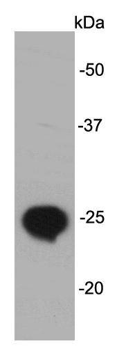
Fig1: Western blot analysis on Lin28B recombinant protein lysates using anti- Lin28B mouse mAb.Application
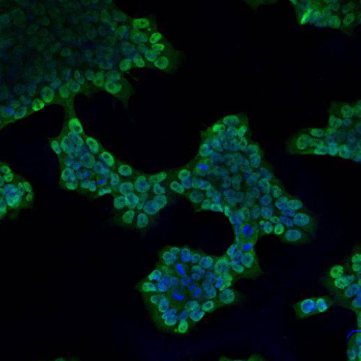
Fig2: ICC staining Lin28B in NCCIT cells (green). Cells were fixed in paraformaldehyde, permeabilised with 0.25% Triton X100/PBS. and counterstained with DAPI in order to highlight the nucleus (blue).| Product Name | Lin28B [A2-4-5] |
|---|---|
| Antibody Type | Primary Antibodies |
| Antigen Alias | CSDD 2 antibody CSDD2 antibody FLJ16517 antibody Lin 28 homolog B (C. elegans) antibody Lin 28.2 antibody Lin-28B antibody LIN28B antibody LN28B_HUMAN antibody Protein lin-28 homolog B antibody |
| Product description | LIN28 is thought to regulate the self-renewal of stem cells. In nematodes, the LIN28 homolog lin-28 is a heterochronic gene that determines the onset of early larval stages of developmental events in Caenorhabditis elegans, by regulating the self-renewal of nematode stem cells in the skin (called seam cells) and vulva (called VPCs) during development. In mice, LIN28 is highly expressed in mouse embryonic stem cells and during early embryogenesis.It acts as a suppressor of microRNA (miRNA) biogenesis by specifically binding the precursor let-7 (pre-let-7), a miRNA precursor. Lin28B is high expression in testis, fetal liver, placenta and in hepatocellular carcinoma (HCC). |
| Immunogen | recombinant protein |
| Clonality | Monoclonal |
|---|---|
| Isotype | IgG2a |
| Host Species | Mouse |
| Tested Applications | WBICCIHC |
| WB:1:100-1:500 ICC:1:100-1:200 |
|
| Species Reactivity | Human |
| Concentration | 2mg/ml |
| Alternative Names | CSDD 2 antibody CSDD2 antibody FLJ16517 antibody Lin 28 homolog B (C. elegans) antibody Lin 28.2 antibody Lin-28B antibody LIN28B antibody LN28B_HUMAN antibody Protein lin-28 homolog B antibody |
|---|---|
| Molecular Weight(MW) | ~27 kDa |
| Cellular Localization | Cytoplasm, nucleus |
| SwissProt ID | Q6ZN17 |
|---|
Application
Fig1: Western blot analysis on Lin28B recombinant protein lysates using anti- Lin28B mouse mAb.
Application
Fig2: ICC staining Lin28B in NCCIT cells (green). Cells were fixed in paraformaldehyde, permeabilised with 0.25% Triton X100/PBS. and counterstained with DAPI in order to highlight the nucleus (blue).| Positive Control | NCCIT, Lin28B recombinant protein |
|---|---|
| Application Notes | WB:1:100-1:500 ICC:1:100-1:200 |
| Form | Liquid |
|---|---|
| Storage Instructions | Store at +4℃ after thawing. Aliquot store at -20℃ or -80℃. Avoid repeated freeze / thaw cycles. |
| Storage Buffer | 1*TBS (pH7.4), 0.5%BSA, 40%Glycerol. Preservative: 0.05% Sodium Azide. |


