Application
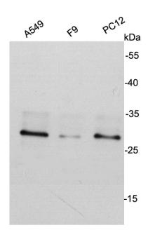
Fig1: Western blot analysis on different cell lysates using anti- CDK2 mouse mAb.Application
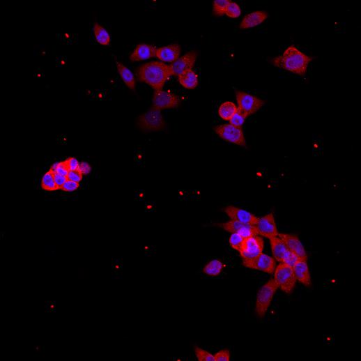
Fig2: ICC staining CDK2 in NCCIT cells (red). Cells were fixed in paraformaldehyde, permeabilised with 0.25% Triton X100/PBS and counterstained with DAPI in order to highlight the nucleus (blue).| Product Name | CDK2 [10-D7] |
|---|---|
| Antibody Type | Primary Antibodies |
| Antigen Alias | Cdc2 related protein kinase antibody cdc2-related protein kinase antibody CDC28 antibody CDC2A antibody Cdk 2 antibody CDK1 antibody CDK2 antibody CDK2_HUMAN antibody CDKN2 antibody Cell devision kinase 2 antibody Cell division protein kinase 2 antibody Cyclin dependent kinase 2 antibody cyclin dependent kinase 2-alpha antibody Cyclin-dependent kinase 2 antibody kinase Cdc2 antibody MPF antibody p33 protein kinase antibody p33(CDK2) antibody |
| Product description | Cyclin-dependent kinase 2 (p33CDK2) is an important component of the cell cycle machinery. It acts at the G1-S transition to promote the E2F transcriptional program and the initiation of DNA synthesis, and modulates G2 progression; controls the timing of entry into mitosis/meiosis by controlling the subsequent activation of cyclin B/CDK1 by phosphorylation, and coordinates the activation of cyclin B/CDK1 at the centrosome and in the nucleus. Inhibition of CDK2-cyclin complexes can also be attributed to association with p27 Kip1 and p21 Waf1/Cip1. |
| Immunogen | peptide |
| Clonality | Monoclonal |
|---|---|
| Isotype | IgG2b |
| Host Species | Mouse |
| Tested Applications | WBICCIHCFC |
| WB:1:2000 ICC:1:500 |
|
| Species Reactivity | HumanMouseRat |
| Concentration | 2mg/ml |
| Alternative Names | Cdc2 related protein kinase antibody cdc2-related protein kinase antibody CDC28 antibody CDC2A antibody Cdk 2 antibody CDK1 antibody CDK2 antibody CDK2_HUMAN antibody CDKN2 antibody Cell devision kinase 2 antibody Cell division protein kinase 2 antibody Cyclin dependent kinase 2 antibody cyclin dependent kinase 2-alpha antibody Cyclin-dependent kinase 2 antibody kinase Cdc2 antibody MPF antibody p33 protein kinase antibody p33(CDK2) antibody |
|---|---|
| Molecular Weight(MW) | ~33kDa |
| Cellular Localization | Cytoplasm, nucleus |
| SwissProt ID | P24941 |
|---|
Application
Fig1: Western blot analysis on different cell lysates using anti- CDK2 mouse mAb.
Application
Fig2: ICC staining CDK2 in NCCIT cells (red). Cells were fixed in paraformaldehyde, permeabilised with 0.25% Triton X100/PBS and counterstained with DAPI in order to highlight the nucleus (blue).| Positive Control | A549, F9, PC12, NCCIT |
|---|---|
| Application Notes | WB:1:2000 ICC:1:500 |
| Form | Liquid |
|---|---|
| Storage Instructions | Store at +4℃ after thawing. Aliquot store at -20℃ or -80℃. Avoid repeated freeze / thaw cycles. |
| Storage Buffer | 1*TBS (pH7.4), 1%BSA, 40%Glycerol. Preservative: 0.05% Sodium Azide. |


