Application
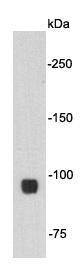
Fig1: Western blot analysis on 293T cell lysates using anti- Beta-catenin mouse mAb.Application
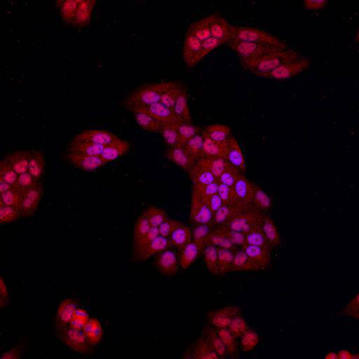
Fig2: ICC staining Beta-catenin in Hela cells (red). Cells were fixed in paraformaldehyde, permeabilised with 0.25% Triton X100/PBS and counterstained with DAPI in order to highlight the nucleus (blue).| Product Name | Beta-catenin [10-C0-B7] |
|---|---|
| Antibody Type | Primary Antibodies |
| Antigen Alias | Beta catenin antibody Beta-catenin antibody Cadherin associated protein antibody Catenin (cadherin associated protein), beta 1, 88kDa antibody Catenin beta 1 antibody Catenin beta-1 antibody CATNB antibody CHBCAT antibody CTNB1_HUMAN antibody CTNNB antibody CTNNB1 antibody DKFZp686D02253 antibody FLJ25606 antibody FLJ37923 antibody OTTHUMP00000162082 antibody OTTHUMP00000165222 antibody OTTHUMP00000165223 antibody OTTHUMP00000209288 antibody OTTHUMP00000209289 antibody |
| Product description | Catenin (cadherin-associated protein), beta 1, 88kDa, also called beta-catenin (or β-catenin), is a dual function protein, regulating the coordination of cell–cell adhesion and gene transcription. The cellular level of beta-catenin is mostly controlled by its ubiquitinylation and proteosomal degradation. Cell–cell adhesion complexes are essential for the formation of complex animal tissues. β-catenin is part of a protein complex that form the so-called adherens junctions. Beta-catenin has a central role in directing several developmental processes, as it can directly bind transcription factors and be regulated by a diffusible extracellular substance: Wnt. It acts upon early embryos to induce entire body regions, as well as individual cells in later stages of development. It also regulates physiological regeneration processes. Wnt signaling and beta-catenin dependent gene expression plays a critical role during the formation of different body regions in the early embryo. |
| Immunogen | peptide |
| Clonality | Monoclonal |
|---|---|
| Isotype | IgG2b |
| Host Species | Mouse |
| Tested Applications | WBICCIHCFC |
| WB:1:2000 ICC:1:200 |
|
| Species Reactivity | HumanMouseRat |
| Concentration | 2mg/ml |
| Alternative Names | Beta catenin antibody
Beta-catenin antibody
Cadherin associated protein antibody
Catenin (cadherin associated protein) beta 1 88kDa antibody Catenin beta 1 antibody Catenin beta-1 antibody CATNB antibody CHBCAT antibody CTNB1_HUMAN antibody CTNNB antibody CTNNB1 antibody DKFZp686D02253 antibody FLJ25606 antibody FLJ37923 antibody OTTHUMP00000162082 antibody OTTHUMP00000165222 antibody OTTHUMP00000165223 antibody OTTHUMP00000209288 antibody OTTHUMP00000209289 antibody |
|---|---|
| Molecular Weight(MW) | 85kDa |
| Cellular Localization | Cytoplasm, nucleus |
| SwissProt ID | P35222 |
|---|
Application
Fig1: Western blot analysis on 293T cell lysates using anti- Beta-catenin mouse mAb.
Application
Fig2: ICC staining Beta-catenin in Hela cells (red). Cells were fixed in paraformaldehyde, permeabilised with 0.25% Triton X100/PBS and counterstained with DAPI in order to highlight the nucleus (blue).| Positive Control | Hela, 293T |
|---|---|
| Application Notes | WB:1:2000 ICC:1:200 |
| Form | Liquid |
|---|---|
| Storage Instructions | Store at +4℃ after thawing. Aliquot store at -20℃ or -80℃. Avoid repeated freeze / thaw cycles. |
| Storage Buffer | 1*TBS (pH7.4), 1%BSA, 40%Glycerol. Preservative: 0.05% Sodium Azide. |


