Application
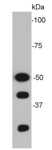
Fig1: Western blot analysis of NF-κB p105/p50 on MCF-7 cell lysates.Application
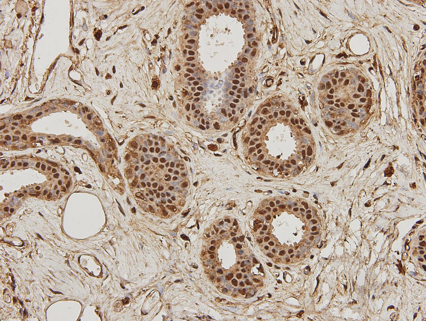
Fig2: Immunohistochemical analysis of paraffin-embedded human breast carcinoma tissue using anti-NF-κB p105/p50 antibody. Counter stained with hematoxylin.Application
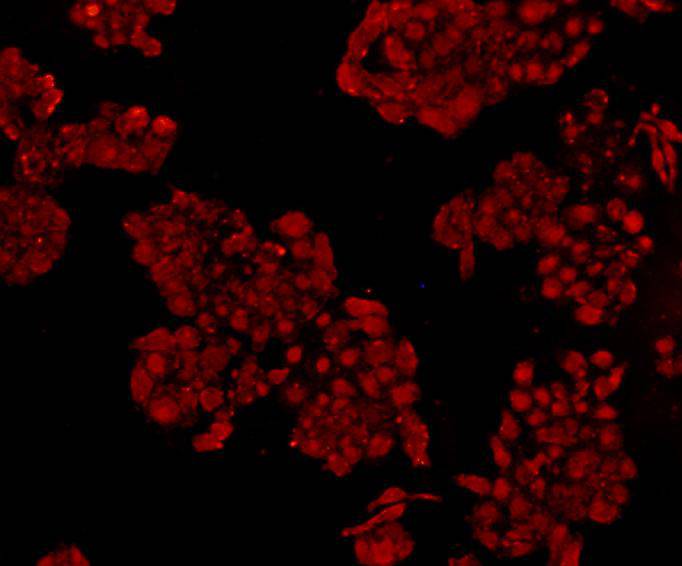
Fig3: ICC staining NF-κB p105/p50 in HepG2 cells (red). Cells were fixed in paraformaldehyde, permeabilised with 0.25% Triton X100/PBS.| Product Name | NF-κB p105/p50 [A2-7] |
|---|---|
| Antibody Type | Primary Antibodies |
| Antigen Alias | DKFZp686C01211 antibody DNA binding factor KBF1 antibody DNA binding factor KBF1 EBP1 antibody DNA-binding factor KBF1 antibody EBP 1 antibody EBP-1 antibody EBP1 antibody KBF1 antibody MGC54151 antibody NF kappa B antibody NF kappaB antibody NF kappabeta antibody NF kB1 antibody NFkappaB antibody NFKB 1 antibody NFKB p105 antibody NFKB p50 antibody Nfkb1 antibody NFKB1_HUMAN antibody Nuclear factor kappa B DNA binding subunit antibody Nuclear factor kappa-B, subunit 1 antibody Nuclear factor NF kappa B p105 subunit antibody Nuclear factor NF kappa B p50 subunit antibody Nuclear factor NF-kappa-B p50 subunit antibody Nuclear factor of kappa light chain gene enhancer in B cells 1 antibody Nuclear factor of kappa light polypeptide gene enhancer in B cells 1 antibody Nuclear factor of kappa light polypeptide gene enhancer in B-cells 1 antibody p105 antibody p50 antibody p84/NF-kappa-B1 p98 antibody Transcription factor NFKB1 antibody |
| Product description | NF-kappa-B is a pleiotropic transcription factor present in almost all cell types and is the endpoint of a series of signal transduction events that are initiated by a vast array of stimuli related to many biological processes such as inflammation, immunity, differentiation, cell growth, tumorigenesis and apoptosis. NF-kappa-B is a homo- or heterodimeric complex formed by the Rel-like domain-containing proteins RELA/p65, RELB, NFKB1/p105, NFKB1/p50, REL and NFKB2/p52 and the heterodimeric p65-p50 complex appears to be most abundant one. The dimers bind at kappa-B sites in the DNA of their target genes and the individual dimers have distinct preferences for different kappa-B sites that they can bind with distinguishable affinity and specificity. Different dimer combinations act as transcriptional activators or repressors, respectively. NF-kappa-B is controlled by various mechanisms of post-translational modification and subcellular compartmentalization as well as by interactions with other cofactors or corepressors. NF-kappa-B heterodimeric p65-p50 and RelB-p50 complexes are transcriptional activators. |
| Immunogen | This antibody is produced by immunizing mice with a synthetic peptide (KLH-coupled) corresponding to NF-κB p105/p50. |
| Clonality | Monoclonal |
|---|---|
| Isotype | IgM |
| Host Species | Mouse |
| Tested Applications | WBICCIHC |
| WB:1:1,000-1:2,000 ICC:1:200 IHC:1:200-1:500 |
|
| Species Reactivity | HumanMouse |
| Concentration | 2mg/ml |
| Alternative Names | DKFZp686C01211 antibody
DNA binding factor KBF1 antibody
DNA binding factor KBF1 EBP1 antibody
DNA-binding factor KBF1 antibody
EBP 1 antibody
EBP-1 antibody
EBP1 antibody
KBF1 antibody
MGC54151 antibody
NF kappa B antibody
NF kappaB antibody
NF kappabeta antibody
NF kB1 antibody
NFkappaB antibody
NFKB 1 antibody
NFKB p105 antibody
NFKB p50 antibody
Nfkb1 antibody
NFKB1_HUMAN antibody
Nuclear factor kappa B DNA binding subunit antibody
Nuclear factor kappa-B subunit 1 antibody Nuclear factor NF kappa B p105 subunit antibody Nuclear factor NF kappa B p50 subunit antibody Nuclear factor NF-kappa-B p50 subunit antibody Nuclear factor of kappa light chain gene enhancer in B cells 1 antibody Nuclear factor of kappa light polypeptide gene enhancer in B cells 1 antibody Nuclear factor of kappa light polypeptide gene enhancer in B-cells 1 antibody p105 antibody p50 antibody p84/NF-kappa-B1 p98 antibody Transcription factor NFKB1 antibody |
|---|---|
| Molecular Weight(MW) | 50kDa |
| Cellular Localization | Nucleus. Cytoplasm. |
| SwissProt ID | P19838 |
|---|
Application
Fig1: Western blot analysis of NF-κB p105/p50 on MCF-7 cell lysates.
Application
Fig2: Immunohistochemical analysis of paraffin-embedded human breast carcinoma tissue using anti-NF-κB p105/p50 antibody. Counter stained with hematoxylin.
Application
Fig3: ICC staining NF-κB p105/p50 in HepG2 cells (red). Cells were fixed in paraformaldehyde, permeabilised with 0.25% Triton X100/PBS.| Positive Control | MCF-7, HepG2, human breast carcinoma tissue |
|---|---|
| Application Notes | WB:1:1,000-1:2,000 ICC:1:200 IHC:1:200-1:500 |
| Form | Liquid |
|---|---|
| Storage Instructions | Store at +4℃ after thawing. Aliquot store at -20℃. Avoid repeated freeze / thaw cycles. |
| Storage Buffer | 1*TBS (pH7.4), 1%BSA, 40%Glycerol. Preservative: 0.05% Sodium Azide. |


