Application
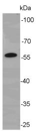
Fig1: Western blot analysis of Cyclin B1 on K562 lysate. Proteins were transferred to a PVDF membrane and blocked with 5% BSA in PBS for 1 hour at room temperature. The primary antibody was used at a 1:500 dilution in 5% BSA at room temperature for 2 hours. Goat Anti-Mouse IgG - HRP Secondary Antibody (HA1006) at 1:5,000 dilution was used for 1 hour at room temperature.Application
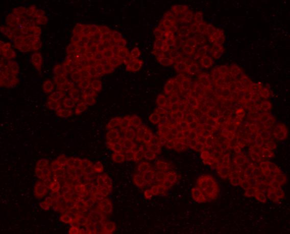
Fig2: ICC staining Cyclin B1 in HepG2 cells (red). Formalin fixed cells were permeabilized with 0.1% Triton X-100 in TBS for 10 minutes at room temperature and blocked with 1% Blocker BSA for 15 minutes at room temperature. Cells were probed with Cyclin B1 monoclonal antibody at a dilution of 1:200 for 1 hour at room temperature, washed with PBS. AlexaFluor®488 Goat anti-Mouse IgG was used as the secondary antibody at 1/100 dilution.Application
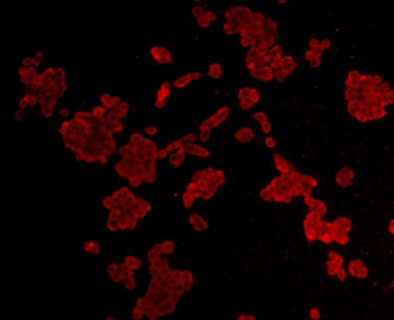
Fig3: ICC staining Cyclin B1 in LOVO cells (red). Formalin fixed cells were permeabilized with 0.1% Triton X-100 in TBS for 10 minutes at room temperature and blocked with 1% Blocker BSA for 15 minutes at room temperature. Cells were probed with Cyclin B1 monoclonal antibody at a dilution of 1:200 for 1 hour at room temperature, washed with PBS. AlexaFluor®488 Goat anti-Mouse IgG was used as the secondary antibody at 1/100 dilution.| Product Name | Cyclin B1 [C2-F2] |
|---|---|
| Antibody Type | Primary Antibodies |
| Antigen Alias | CCNB 1 antibody CCNB antibody ccnb1 antibody CCNB1_HUMAN antibody Cyclin B1 antibody G2 mitotic specific cyclin B1 antibody G2/mitotic-specific cyclin-B1 antibody |
| Product description | Mitogen-activated protein kinases (MAPKs) are a widely conserved family of serine/threonine protein kinases involved in many cellular programs, such as cell proliferation, differentiation, motility, and death. The p44/42 MAPK (Erk1/2) signaling pathway can be activated in response to a diverse range of extracellular stimuli including mitogens, growth factors, and cytokines , and research investigators consider it an important target in the diagnosis and treatment of cancer . Upon stimulation, a sequential three-part protein kinase cascade is initiated, consisting of a MAP kinase kinase kinase (MAPKKK or MAP3K), a MAP kinase kinase (MAPKK or MAP2K), and a MAP kinase (MAPK). Multiple p44/42 MAP3Ks have been identified, including members of the Raf family, as well as Mos and Tpl2/COT. MEK1 and MEK2 are the primary MAPKKs in this pathway. |
| Immunogen | This antibody is produced by immunizing mice with a synthetic peptide (KLH-coupled) corresponding to a region of Cyclin B1. |
| Clonality | Monoclonal |
|---|---|
| Isotype | IgG1 |
| Host Species | Mouse |
| Tested Applications | WBICC |
| WB:1:500: ICC:1:200-1:500: |
|
| Species Reactivity | Human |
| Concentration | 2mg/ml |
| Alternative Names | CCNB 1 antibody CCNB antibody ccnb1 antibody CCNB1_HUMAN antibody Cyclin B1 antibody G2 mitotic specific cyclin B1 antibody G2/mitotic-specific cyclin-B1 antibody |
|---|---|
| Molecular Weight(MW) | 58 kDa (predicted molecular weight: 48 kDa) |
| Cellular Localization | Cytoplasm, Nucleus. |
| SwissProt ID | P14635 |
|---|
Application
Fig1: Western blot analysis of Cyclin B1 on K562 lysate. Proteins were transferred to a PVDF membrane and blocked with 5% BSA in PBS for 1 hour at room temperature. The primary antibody was used at a 1:500 dilution in 5% BSA at room temperature for 2 hours. Goat Anti-Mouse IgG - HRP Secondary Antibody (HA1006) at 1:5,000 dilution was used for 1 hour at room temperature.
Application
Fig2: ICC staining Cyclin B1 in HepG2 cells (red). Formalin fixed cells were permeabilized with 0.1% Triton X-100 in TBS for 10 minutes at room temperature and blocked with 1% Blocker BSA for 15 minutes at room temperature. Cells were probed with Cyclin B1 monoclonal antibody at a dilution of 1:200 for 1 hour at room temperature, washed with PBS. AlexaFluor®488 Goat anti-Mouse IgG was used as the secondary antibody at 1/100 dilution.
Application
Fig3: ICC staining Cyclin B1 in LOVO cells (red). Formalin fixed cells were permeabilized with 0.1% Triton X-100 in TBS for 10 minutes at room temperature and blocked with 1% Blocker BSA for 15 minutes at room temperature. Cells were probed with Cyclin B1 monoclonal antibody at a dilution of 1:200 for 1 hour at room temperature, washed with PBS. AlexaFluor®488 Goat anti-Mouse IgG was used as the secondary antibody at 1/100 dilution.| Positive Control | K562, HepG2, LOVO. |
|---|---|
| Application Notes | WB:1:500: ICC:1:200-1:500: |
| Form | Liquid |
|---|---|
| Storage Instructions | Store at +4℃ after thawing. Aliquot store at -20℃. Avoid repeated freeze / thaw cycles. |
| Storage Buffer | 1*TBS (pH7.4), 1%BSA, 40%Glycerol. Preservative: 0.05% Sodium Azide. |


