Application
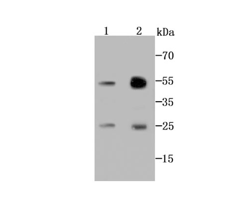
Fig1: Western blot analysis of AK6 on MCF-7 cell (1) and human lung tissue (2) lysate using anti-AK6 antibody at 1/1000 dilution.Application
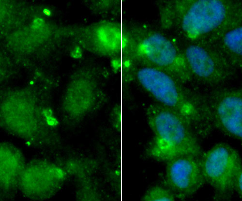
Fig2: ICC staining AK6 in MCF-7 cells (green). The nuclear counter stain is DAPI (blue). Cells were fixed in paraformaldehyde, permeabilised with 0.25% Triton X100/PBS.Application
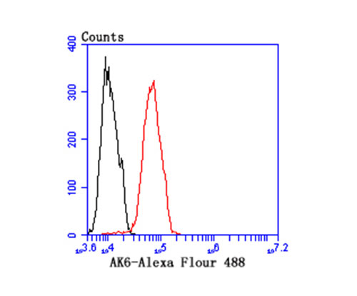
Fig3: Flow cytometric analysis of MCF-7 cells with AK6 antibody at 1/100 dilution (red) compared with an unlabelled control (cells without incubation with primary antibody; black).| Product Name | AK6 [B1-F4] |
|---|---|
| Antibody Type | Primary Antibodies |
| Antigen Alias | AD-004 antibody Adenylate kinase isoenzyme 6 antibody Adrenal gland protein AD-004 antibody AK/ATPase antibody AK6 antibody CGI-137 antibody CIP antibody Coilin-interacting nuclear ATPase protein antibody Dual activity adenylate kinase/ATPase antibody hCINAP antibody KAD6_HUMAN antibody TAF9 antibody |
| Product description | In eukaryotic systems, the process of initiating transcription from protein-coding genes requires the presence of RNA polymerase II and a broad family of auxiliary transcription factors. Such factors can be divided into two major functional classes: the basal factors that mediate the transcription of all Pol II genes, including TFIIA, TFIIB, TFIID, TFIIE, TFIIF and TFIIH, and sequence-specific factors that regulate gene expression. TFIID, one of the basal transcription factors, facilitates the preinitiation complex assembly through direct interactions with the TATA promoter element. TAF9 (transcription initiation factor TFIID subunit 9), also known as TAF9A or TAF II p32, is a 264 amino acid nuclear protein that, is a component of the TFIID complex and is required for cell viability. |
| Immunogen | Peptide |
| Clonality | Monoclonal |
|---|---|
| Isotype | IgG1 |
| Host Species | Mouse |
| Tested Applications | WBICCFC |
| WB:1:250~1:1000 ICC:1:100~1:200 FC:1:100~1:200 Notes:Optimal dilutions/concentrations should be determined by the researcher. |
|
| Species Reactivity | HumanMouseRat |
| Concentration | 2mg/ml |
| Alternative Names | AD-004 antibody Adenylate kinase isoenzyme 6 antibody Adrenal gland protein AD-004 antibody AK/ATPase antibody AK6 antibody CGI-137 antibody CIP antibody Coilin-interacting nuclear ATPase protein antibody Dual activity adenylate kinase/ATPase antibody hCINAP antibody KAD6_HUMAN antibody TAF9 antibody |
|---|---|
| Molecular Weight(MW) | 20 kDa |
| Cellular Localization | Nucleus. |
| SwissProt ID | Q9Y3D8 |
|---|
Application
Fig1: Western blot analysis of AK6 on MCF-7 cell (1) and human lung tissue (2) lysate using anti-AK6 antibody at 1/1000 dilution.
Application
Fig2: ICC staining AK6 in MCF-7 cells (green). The nuclear counter stain is DAPI (blue). Cells were fixed in paraformaldehyde, permeabilised with 0.25% Triton X100/PBS.
Application
Fig3: Flow cytometric analysis of MCF-7 cells with AK6 antibody at 1/100 dilution (red) compared with an unlabelled control (cells without incubation with primary antibody; black).| Positive Control | Human lung tissue lysate, MCF-7. |
|---|---|
| Application Notes | WB:1:250~1:1000 ICC:1:100~1:200 FC:1:100~1:200 Notes:Optimal dilutions/concentrations should be determined by the researcher. |
| Form | Liquid |
|---|---|
| Storage Instructions | Store at +4℃ after thawing. Aliquot store at -20℃ or -80℃. Avoid repeated freeze / thaw cycles. |
| Storage Buffer | 1*TBS (pH7.4), 1%BSA, 40%Glycerol. Preservative: 0.05% Sodium Azide. |


