WB
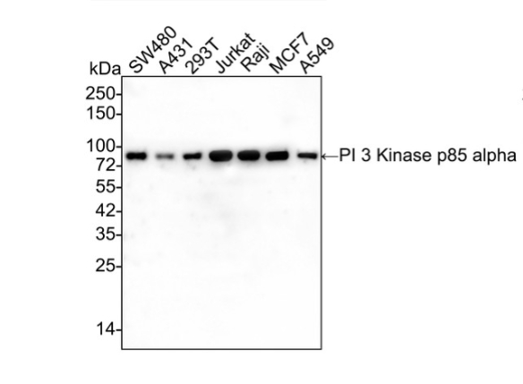
Western blot analysis of PI 3 Kinase p85 alpha on different lysates with Mouse anti-PI 3 Kinase p85 alpha antibody at 1/5,000 dilution. Lane 1: SW480 cell lysate, Lane 2: A431 cell lysate, Lane 3: 293T cell lysate, Lane 4: Jurkat cell lysate, Lane 5: Raji cell lysate, Lane 6: MCF7 cell lysate, Lane 7: A549 cell lysate, Lysates/proteins at 15 µg/Lane. Exposure time: 1 minute 40 seconds; 4-20% SDS-PAGE gel. Proteins were transferred to a PVDF membrane and blocked with 5% NFDM/TBST for 1 hour at room temperature. The primary antibody at 1/5,000 dilution was used in 5% NFDM/TBST at 4℃ overnight. Goat Anti-Mouse IgG - HRP Secondary Antibody at 1/50,000 dilution was used for 1 hour at room temperature.ICC/IF
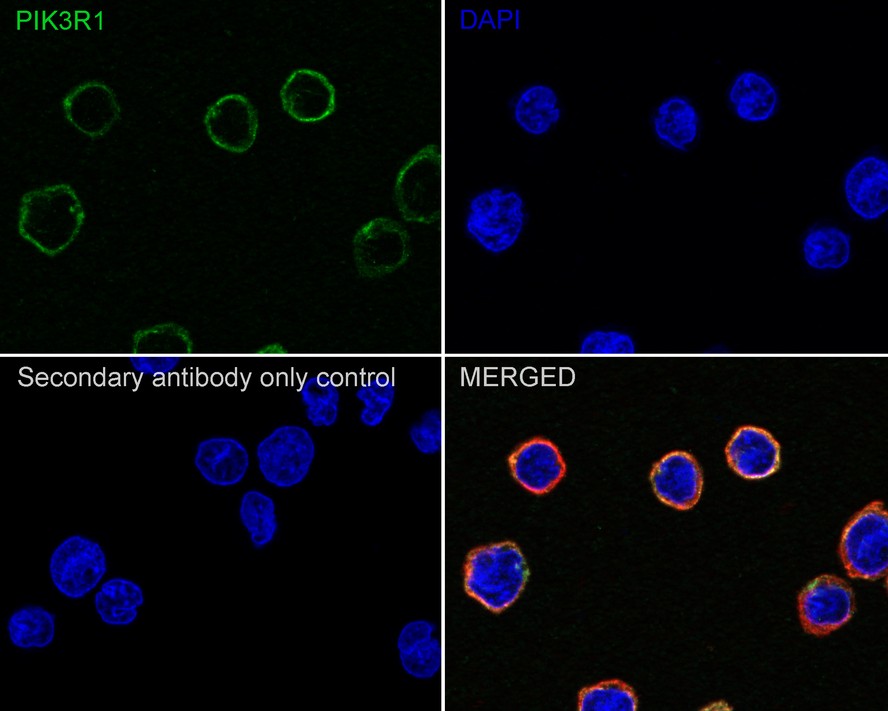
Immunocytochemistry analysis of U-937 cells labeling PI 3 Kinase p85 alpha with Mouse anti-PI 3 Kinase p85 alpha antibody at 1/100 dilution. Cells were fixed in 4% paraformaldehyde for 20 minutes at room temperature, permeabilized with 0.1% Triton X-100 in PBS for 5 minutes at room temperature, then blocked with 1% BSA in 10% negative goat serum for 1 hour at room temperature. Cells were then incubated with Mouse anti-PI 3 Kinase p85 alpha antibody at 1/100 dilution in 1% BSA in PBST overnight at 4 ℃. Goat Anti-Mouse IgG H&L (488) was used as the secondary antibody at 1/1,000 dilution. PBS instead of the primary antibody was used as the secondary antibody only control. Nuclear DNA was labelled in blue with DAPI. beta Tubulin (red) was stained at 1/100 dilution overnight at +4℃. Goat Anti-Rabbit IgG H&L (594) were used as the secondary antibody at 1/1,000 dilution.| Product Name | PI 3 Kinase p85 alpha Recombinant Mouse Monoclonal Antibody |
|---|---|
| Antibody Type | Primary Antibodies |
| Product description | Phosphatidylinositol 3-kinase (PI 3-kinase) is composed of (p85) and (p110) subunits. p85 lacks PI 3-kinase activity and acts as an adapter, coupling p110 to activated protein tyrosine kinase. Two forms of p85 have been described (p85α and p85β), each possessing one SH3 and two SH2 domains. Various p110 isoforms have been identified. p110α and p110β interact with p85α, and p110α has also been shown to interact with p85β in vitro. p110δ expression is restricted to white blood cells. It has been shown to bind p85α and γ, but it apparently does not phosphorylate these subunits. p110δ seems to have the capacity to autophosphorylate. p110γ does not interact with the p85 subunits. It has been shown to be activated by α and βγ heterotrimeric G proteins. |
| Immunogen | Recombinant protein within Human PI3-kinase p85 subunit alpha aa 19-219 / 724. |
| Clonality | monoclonal |
|---|---|
| Isotype | IgG1 |
| Host Species | Mouse |
| Tested Applications | ICC/IFWB |
| WB:1:2000-1:5000 ICC/IF:1:100 |
|
| Species Reactivity | Human |
| Concentration | 1mg/ml |
| Purification | Protein A |
| Alternative Names | GRB1 antibody
p85 alpha antibody
p85 antibody
P85A_HUMAN antibody
Phosphatidylinositol 3 kinase associated p 85 alpha antibody
Phosphatidylinositol 3 kinase regulatory 1 antibody
Phosphatidylinositol 3 kinase regulatory subunit polypeptide 1 (p85 alpha) antibody Phosphatidylinositol 3-kinase 85 kDa regulatory subunit alpha antibody Phosphatidylinositol 3-kinase regulatory subunit alpha antibody Phosphoinositide 3 kinase regulatory subunit 1 (alpha) antibody PI3 kinase p85 subunit alpha antibody PI3-kinase regulatory subunit alpha antibody PI3-kinase subunit p85-alpha antibody PI3K antibody PI3K regulatory subunit alpha antibody Pik3r1 antibody PtdIns 3 kinase p85 alpha antibody PtdIns-3-kinase regulatory subunit alpha antibody PtdIns-3-kinase regulatory subunit p85-alpha antibody |
|---|---|
| Molecular Weight(MW) | 84kDa |
| Cellular Localization | Cytosol, Membrane, Nucleus. |
| SwissProt ID | P27986 |
|---|
WB
Western blot analysis of PI 3 Kinase p85 alpha on different lysates with Mouse anti-PI 3 Kinase p85 alpha antibody at 1/5,000 dilution. Lane 1: SW480 cell lysate, Lane 2: A431 cell lysate, Lane 3: 293T cell lysate, Lane 4: Jurkat cell lysate, Lane 5: Raji cell lysate, Lane 6: MCF7 cell lysate, Lane 7: A549 cell lysate, Lysates/proteins at 15 µg/Lane. Exposure time: 1 minute 40 seconds; 4-20% SDS-PAGE gel. Proteins were transferred to a PVDF membrane and blocked with 5% NFDM/TBST for 1 hour at room temperature. The primary antibody at 1/5,000 dilution was used in 5% NFDM/TBST at 4℃ overnight. Goat Anti-Mouse IgG - HRP Secondary Antibody at 1/50,000 dilution was used for 1 hour at room temperature.
ICC/IF
Immunocytochemistry analysis of U-937 cells labeling PI 3 Kinase p85 alpha with Mouse anti-PI 3 Kinase p85 alpha antibody at 1/100 dilution. Cells were fixed in 4% paraformaldehyde for 20 minutes at room temperature, permeabilized with 0.1% Triton X-100 in PBS for 5 minutes at room temperature, then blocked with 1% BSA in 10% negative goat serum for 1 hour at room temperature. Cells were then incubated with Mouse anti-PI 3 Kinase p85 alpha antibody at 1/100 dilution in 1% BSA in PBST overnight at 4 ℃. Goat Anti-Mouse IgG H&L (488) was used as the secondary antibody at 1/1,000 dilution. PBS instead of the primary antibody was used as the secondary antibody only control. Nuclear DNA was labelled in blue with DAPI. beta Tubulin (red) was stained at 1/100 dilution overnight at +4℃. Goat Anti-Rabbit IgG H&L (594) were used as the secondary antibody at 1/1,000 dilution.| Application Notes | WB:1:2000-1:5000 ICC/IF:1:100 |
|---|
| Form | Liquid |
|---|---|
| Storage Instructions | Store at +4℃ after thawing. Aliquot store at -20℃ or -80℃. Avoid repeated freeze / thaw cycles. |
| Storage Buffer | 1*TBS (pH7.4), 1%BSA, 40%Glycerol. Preservative: 0.05% Sodium Azide. |


