Application
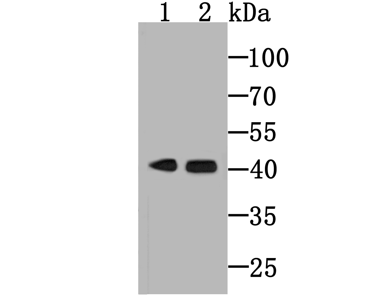
Fig1: Western blot analysis of Haptoglobin on human lung (1) and human liver (2) tissue lysate using anti-Haptoglobin antibody at 1/500 dilution.Application
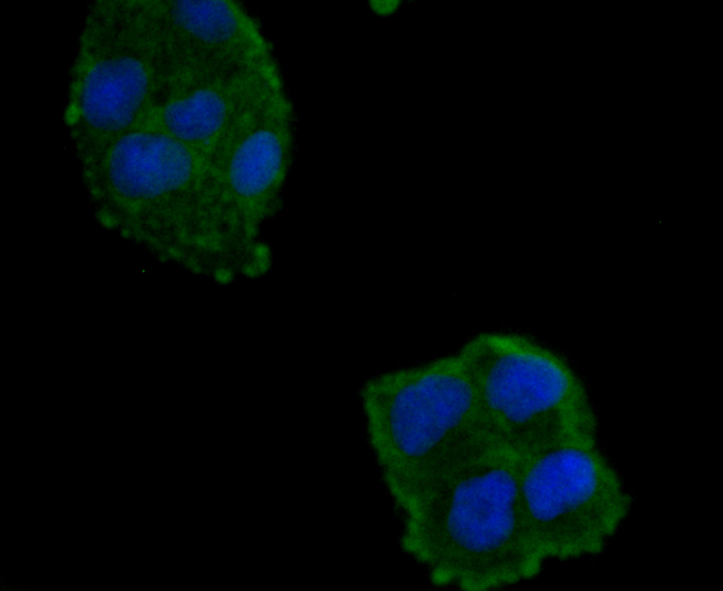
Fig2: ICC staining Haptoglobin (green) in Hela cells. The nuclear counter stain is DAPI (blue). Cells were fixed in paraformaldehyde, permeabilised with 0.25% Triton X100/PBS.Application
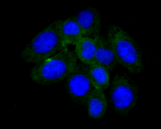
Fig3: ICC staining Haptoglobin (green) in HepG2 cells. The nuclear counter stain is DAPI (blue). Cells were fixed in paraformaldehyde, permeabilised with 0.25% Triton X100/PBS.Application
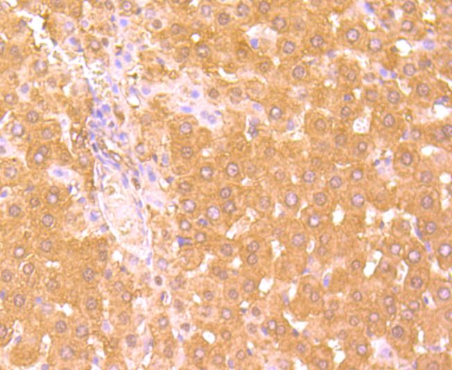
Fig4: Immunohistochemical analysis of paraffin-embedded rat liver tissue using anti-Haptoglobin antibody. Counter stained with hematoxylin.Application
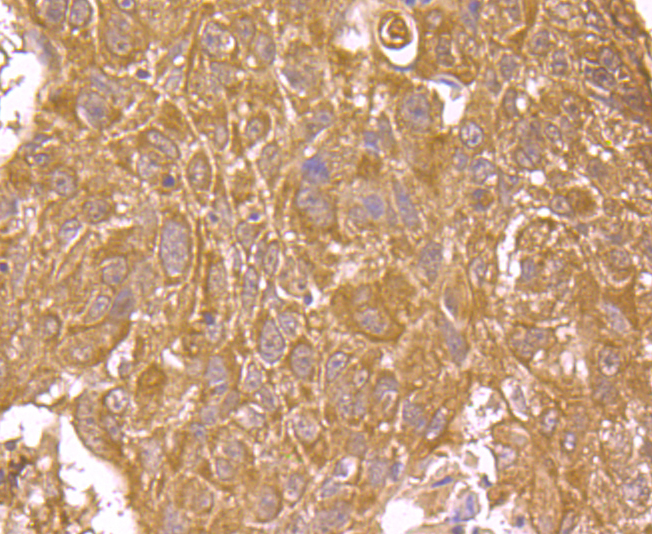
Fig5: Immunohistochemical analysis of paraffin-embedded human lung cancer tissue using anti-Haptoglobin antibody. Counter stained withApplication
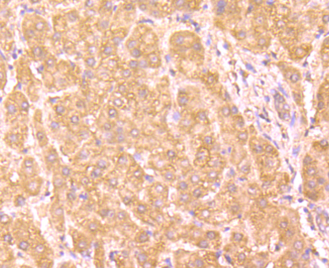
Fig6: Immunohistochemical analysis of paraffin-embedded human liver tissue using anti-Haptoglobin antibody. Counter stained with hematoxylin.Application
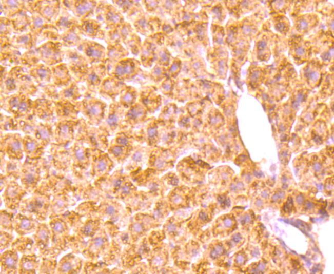
Fig7: Immunohistochemical analysis of paraffin-embedded mouse liver tissue using anti-Haptoglobin antibody. Counter stained with hematoxylin.| Product Name | Haptoglobin [5-D3] |
|---|---|
| Antibody Type | Primary Antibodies |
| Antigen Alias | Binding peptide antibody BP antibody Haptoglobin alpha chain antibody Haptoglobin alpha(1S) beta antibody Haptoglobin alpha(2FS) beta antibody Haptoglobin beta chain antibody Haptoglobin, alpha polypeptide antibody Haptoglobin, beta polypeptide antibody HP antibody HP2 ALPHA2 antibody HP2ALPHA2 antibody HPA1S antibody HPT antibody HPT_HUMAN antibody MGC111141 antibody Zonulin antibody |
| Product description | As a result of hemolysis, hemoglobin is found to accumulate in the kidney and is secreted in the urine. Haptoglobin captures, and combines with free plasma hemoglobin to allow hepatic recycling of heme iron and to prevent kidney damage. Haptoglobin also acts as an Antimicrobial; Antioxidant, has antibacterial activity and plays a role in modulating many aspects of the acute phase response. Hemoglobin/haptoglobin complexes are rapidely cleared by the macrophage CD163 scavenger receptor expressed on the surface of liver Kupfer cells through an endocytic lysosomal degradation pathway. Uncleaved haptoglogin, also known as zonulin, plays a role in intestinal permeability, allowing intercellular tight junction disassembly, and controlling the equilibrium between tolerance and immunity to non-self antigens. |
| Immunogen | Native protein |
| Clonality | Monoclonal |
|---|---|
| Isotype | IgG1 |
| Host Species | Mouse |
| Tested Applications | WBIHCICC |
| WB:1:500-1:2,000 ICC:1:100-1:500 IHC:1:50-1:200 |
|
| Species Reactivity | HumanMouseRat |
| Concentration | 2mg/ml |
| Alternative Names | Binding peptide antibody
BP antibody
Haptoglobin alpha chain antibody
Haptoglobin alpha(1S) beta antibody
Haptoglobin alpha(2FS) beta antibody
Haptoglobin beta chain antibody
Haptoglobin alpha polypeptide antibody Haptoglobin beta polypeptide antibody HP antibody HP2 ALPHA2 antibody HP2ALPHA2 antibody HPA1S antibody HPT antibody HPT_HUMAN antibody MGC111141 antibody Zonulin antibody |
|---|---|
| Molecular Weight(MW) | 42 kDa |
| Cellular Localization | Extracellular region, secreted. |
| SwissProt ID | P00738 |
|---|
Application
Fig1: Western blot analysis of Haptoglobin on human lung (1) and human liver (2) tissue lysate using anti-Haptoglobin antibody at 1/500 dilution.
Application
Fig2: ICC staining Haptoglobin (green) in Hela cells. The nuclear counter stain is DAPI (blue). Cells were fixed in paraformaldehyde, permeabilised with 0.25% Triton X100/PBS.
Application
Fig3: ICC staining Haptoglobin (green) in HepG2 cells. The nuclear counter stain is DAPI (blue). Cells were fixed in paraformaldehyde, permeabilised with 0.25% Triton X100/PBS.
Application
Fig4: Immunohistochemical analysis of paraffin-embedded rat liver tissue using anti-Haptoglobin antibody. Counter stained with hematoxylin.
Application
Fig5: Immunohistochemical analysis of paraffin-embedded human lung cancer tissue using anti-Haptoglobin antibody. Counter stained with
Application
Fig6: Immunohistochemical analysis of paraffin-embedded human liver tissue using anti-Haptoglobin antibody. Counter stained with hematoxylin.
Application
Fig7: Immunohistochemical analysis of paraffin-embedded mouse liver tissue using anti-Haptoglobin antibody. Counter stained with hematoxylin.| Positive Control | Human lung and human liver tissue lysate, HepG2, Hela, rat liver tissue, mouse liver tissue, human liver tissue, human lung cancer tissue. |
|---|---|
| Application Notes | WB:1:500-1:2,000 ICC:1:100-1:500 IHC:1:50-1:200 |
| Form | Liquid |
|---|---|
| Storage Instructions | Store at +4℃after thawing. Aliquot store at -20℃or -80℃. Avoid repeated freeze / thaw cycles. |
| Storage Buffer | 1*TBS (pH7.4), 1%BSA, 40%Glycerol. Preservative: 0.05% Sodium Azide. |


