WB
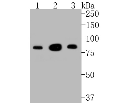
Western blot analysis of PSD95 on different lysates. Proteins were transferred to a PVDF membrane and blocked with 5% BSA in PBS for 1 hour at room temperature. The primary antibody (1/2,000) was used in 5% BSA at room temperature for 2 hours. Goat Anti-Mouse IgG - HRP Secondary Antibody at 1:5,000 dilution was used for 1 hour at room temperature. Positive control: Lane 1: SHG-44 cell lysate, Lane 2: A172 cell lysate, Lane 3: N2A cell lysate.ICC/IF
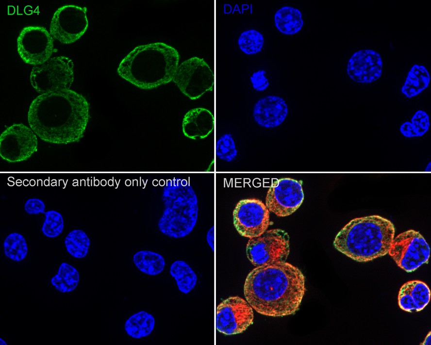
Immunocytochemistry analysis of Neuro-2a cells labeling PSD95 with Mouse anti-PSD95 antibody at 1/100 dilution. Cells were fixed in 4% paraformaldehyde for 15 minutes at room temperature, permeabilized with 0.1% Triton X-100 in PBS for 15 minutes at room temperature, then blocked with 1% BSA in 10% negative goat serum for 1 hour at room temperature. Cells were then incubated with Mouse anti-PSD95 antibody at 1/100 dilution in 1% BSA in PBST overnight at 4 ℃. Goat Anti-Mouse IgG H&L (488) was used as the secondary antibody at 1/1,000 dilution. PBS instead of the primary antibody was used as the secondary antibody only control. Nuclear DNA was labelled in blue with DAPI. beta Tubulin (red) was stained at 1/100 dilution overnight at +4℃. Goat Anti-Rabbit IgG H&L (594) were used as the secondary antibody at 1/1,000 dilution.FC
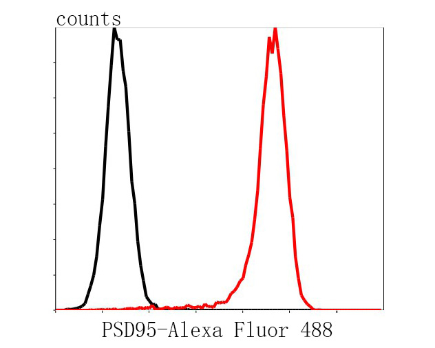
Flow cytometric analysis of PSD95 was done on Hela cells. The cells were fixed, permeabilized and stained with the primary antibody (1/100) (red). After incubation of the primary antibody at room temperature for an hour, the cells were stained with a Alexa Fluor 488-conjugated Goat anti-Mouse IgG Secondary antibody at 1/1000 dilution for 30 minutes.Unlabelled sample was used as a control (cells without incubation with primary antibody; black).| Product Name | PSD95 Mouse Monoclonal Antibody |
|---|---|
| Antibody Type | Primary Antibodies |
| Immunogen | Synthetic peptide within N-terminal human PSD95. |
| Clonality | Monoclonal |
|---|---|
| Isotype | IgG2b |
| Host Species | Mouse |
| Tested Applications | FCICC/IFWB |
| WB:1:2000-1:5000 ICC/IF:1:100 FC:1:50-1:100 |
|
| Species Reactivity | HumanMouseRat |
| Concentration | 1mg/ml |
| Purification | Affinity purified |
| Molecular Weight(MW) | 80kDa |
|---|---|
| Cellular Localization | Cell membrane, postsynaptic density, synapse, Cytoplasm, axon, dendritic spine, dendrite, presynapse. |
| SwissProt ID | P78352 |
|---|
WB
Western blot analysis of PSD95 on different lysates. Proteins were transferred to a PVDF membrane and blocked with 5% BSA in PBS for 1 hour at room temperature. The primary antibody (1/2,000) was used in 5% BSA at room temperature for 2 hours. Goat Anti-Mouse IgG - HRP Secondary Antibody at 1:5,000 dilution was used for 1 hour at room temperature. Positive control: Lane 1: SHG-44 cell lysate, Lane 2: A172 cell lysate, Lane 3: N2A cell lysate.
ICC/IF
Immunocytochemistry analysis of Neuro-2a cells labeling PSD95 with Mouse anti-PSD95 antibody at 1/100 dilution. Cells were fixed in 4% paraformaldehyde for 15 minutes at room temperature, permeabilized with 0.1% Triton X-100 in PBS for 15 minutes at room temperature, then blocked with 1% BSA in 10% negative goat serum for 1 hour at room temperature. Cells were then incubated with Mouse anti-PSD95 antibody at 1/100 dilution in 1% BSA in PBST overnight at 4 ℃. Goat Anti-Mouse IgG H&L (488) was used as the secondary antibody at 1/1,000 dilution. PBS instead of the primary antibody was used as the secondary antibody only control. Nuclear DNA was labelled in blue with DAPI. beta Tubulin (red) was stained at 1/100 dilution overnight at +4℃. Goat Anti-Rabbit IgG H&L (594) were used as the secondary antibody at 1/1,000 dilution.
FC
Flow cytometric analysis of PSD95 was done on Hela cells. The cells were fixed, permeabilized and stained with the primary antibody (1/100) (red). After incubation of the primary antibody at room temperature for an hour, the cells were stained with a Alexa Fluor 488-conjugated Goat anti-Mouse IgG Secondary antibody at 1/1000 dilution for 30 minutes.Unlabelled sample was used as a control (cells without incubation with primary antibody; black).| Application Notes | WB:1:2000-1:5000 ICC/IF:1:100 FC:1:50-1:100 |
|---|
| Form | Liquid |
|---|---|
| Storage Instructions | Store at +4℃ after thawing. Aliquot store at -20℃ or -80℃. Avoid repeated freeze / thaw cycles. |
| Storage Buffer | 1*TBS (pH7.4), 1%BSA, 40%Glycerol. Preservative: 0.05% Sodium Azide. |


