Application
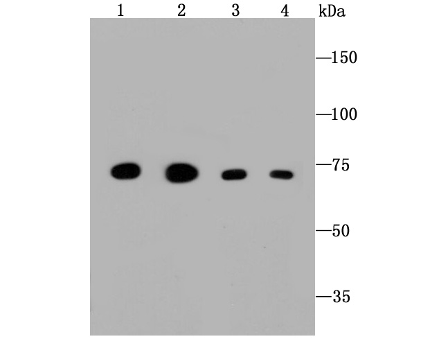
Fig1: Western blot analysis on different cell lysates using anti-Grp75 Mouse mAb. Positive control: Lane 1: HepG2 Lane 2: L929 Lane 3: Hela Lane 4: JurkatApplication
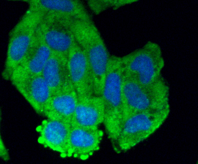
Fig2: Immunocytochemical staining of Hela cells using anti-Grp75 Mouse mAb.Application
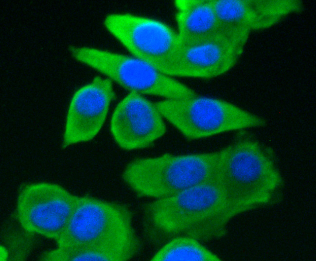
Fig3: Immunocytochemical staining of PC-3M cells using anti-Grp75 Mouse mAb.Application
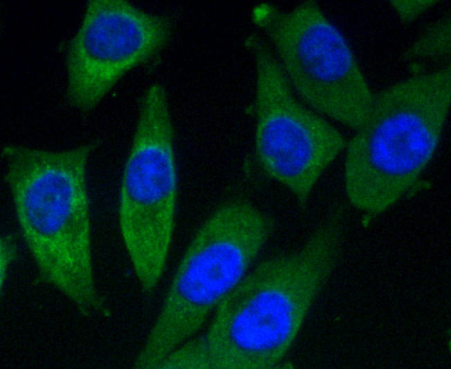
Fig4: Immunocytochemical staining of MCF-7 cells using anti-Grp75 Mouse mAb.Application
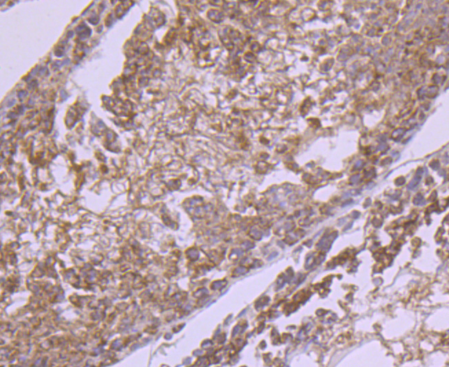
Fig5: Immunohistochemical analysis of paraffin- embedded human breast carcinoma tissue using anti-Grp75 Mouse mAb.Application
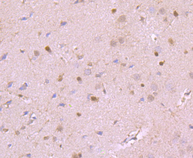
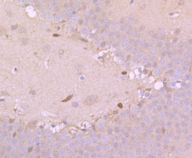
Fig6: Immunohistochemical analysis of paraffin- embedded mouse colon tissue using anti-Grp75 Mouse mAb.Application
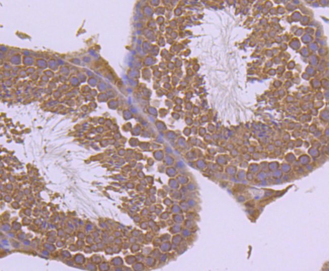
Fig7: Immunohistochemical analysis of paraffin- embedded mouse prostate tissue using anti-Grp75 Mouse mAb.Application
Fig8: Flow cytometric analysis of HepG2 cells with Grp75 antibody at 1/50 dilution (blue) compared with an unlabelled control (cells without incubation with primary antibody; red). Alexa Fluor 488-conjugated Goat anti mouse IgG was used as the secondary antibody.| Product Name | Grp75 [A6-A5-B1] |
|---|---|
| Antibody Type | Primary Antibodies |
| Antigen Alias | 75 kDa glucose regulated protein antibody 75 kDa glucose-regulated protein antibody CSA antibody Glucose Regulated Protein antibody Grp 75 antibody GRP-75 antibody GRP75 antibody GRP75_HUMAN antibody Heat shock 70 kDa protein 9 antibody Heat shock 70kD protein 9 antibody heat shock 70kDa protein 9 antibody Heat shock 70kDa protein 9B antibody Heat shock protein 74 kDa A antibody Heat shock protein A antibody Heat shock protein cognate 74 antibody Hsc74 antibody Hsp74 antibody Hsp74a antibody HSPA9 antibody Hspa9a antibody HSPA9B antibody MGC4500 antibody mitochondrial antibody Mortalin 2 antibody Mortalin antibody Mortalin perinuclear antibody Mortalin2 antibody MOT 2 antibody MOT antibody MOT2 antibody Mthsp70 antibody p66 mortalin antibody P66 MOT antibody PBP74 antibody Peptide binding protein 74 antibody Peptide-binding protein 74 antibody Stress 70 protein mitochondrial antibody Stress 70 protein mitochondrial precursor antibody Stress-70 protein antibody |
| Product description | The HSP 70 family is composed of four highly conserved proteins: HSP 70, HSC 70, GRP 75 and GRP 78. These proteins serve a variety of roles: they act as molecular chaperones facilitating the assembly of multi-protein complexes, participate in the translocation of polypeptides across cell membranes and to the nucleus, and aid in the proper folding of nascent polypeptide chains. All members of the family, except HSP 70, are constitutively expressed in primate cells. HSP 70 expression is strongly induced in response to heat stress. HSP 70 and HSC 70 play key roles in the cytosolic endoplasmic reticulum and mitochondrial import machinery and are found in both the cytosol and nucleus of mammalian cells. Both HSP 70 and HSC 70 are involved in the chaperoning of nascent polypeptide chains and in protecting cells against the accumulation of improperly folded proteins. GRP 78 is localized in the endoplasmic reticulum, where it receives imported secretory proteins and is involved in the folding and translocation of nascent peptide chains. GRP 75 expression is restricted to the mitochondrial matrix and aids in the translocation and folding of nascent polypeptide chains of both nuclear and mitochondrial origin. GRP 75 and GRP 78 are unresponsive to heat stress and are induced by glucose deprivation. It has been postulated that members of the HSP 70 family act as force-generating motors, relying on the hydrolysis of ATP for their activity |
| Immunogen | peptide |
| Clonality | Monoclonal |
|---|---|
| Isotype | IgM |
| Host Species | Mouse |
| Tested Applications | WBICCIHCFC |
| WB:1:1,000-1:2,000 ICC:1:100-1:500 IHC:1:50-1:200 FC:1:50-1:100 |
|
| Species Reactivity | HumanMouseRat |
| Concentration | 2mg/ml |
| Alternative Names | 75 kDa glucose regulated protein antibody 75 kDa glucose-regulated protein antibody CSA antibody Glucose Regulated Protein antibody Grp 75 antibody GRP-75 antibody GRP75 antibody GRP75_HUMAN antibody Heat shock 70 kDa protein 9 antibody Heat shock 70kD protein 9 antibody heat shock 70kDa protein 9 antibody Heat shock 70kDa protein 9B antibody Heat shock protein 74 kDa A antibody Heat shock protein A antibody Heat shock protein cognate 74 antibody Hsc74 antibody Hsp74 antibody Hsp74a antibody HSPA9 antibody Hspa9a antibody HSPA9B antibody MGC4500 antibody mitochondrial antibody Mortalin 2 antibody Mortalin antibody Mortalin perinuclear antibody Mortalin2 antibody MOT 2 antibody MOT antibody MOT2 antibody Mthsp70 antibody p66 mortalin antibody P66 MOT antibody PBP74 antibody Peptide binding protein 74 antibody Peptide-binding protein 74 antibody Stress 70 protein mitochondrial antibody Stress 70 protein mitochondrial precursor antibody Stress-70 protein antibody |
|---|---|
| Molecular Weight(MW) | 74kDa |
| Cellular Localization | Mitochondrion, Nucleus |
| SwissProt ID | P38646 |
|---|
Application
Fig1: Western blot analysis on different cell lysates using anti-Grp75 Mouse mAb. Positive control: Lane 1: HepG2 Lane 2: L929 Lane 3: Hela Lane 4: Jurkat
Application
Fig2: Immunocytochemical staining of Hela cells using anti-Grp75 Mouse mAb.
Application
Fig3: Immunocytochemical staining of PC-3M cells using anti-Grp75 Mouse mAb.
Application
Fig4: Immunocytochemical staining of MCF-7 cells using anti-Grp75 Mouse mAb.
Application
Fig5: Immunohistochemical analysis of paraffin- embedded human breast carcinoma tissue using anti-Grp75 Mouse mAb.
Application
Fig6: Immunohistochemical analysis of paraffin- embedded mouse colon tissue using anti-Grp75 Mouse mAb.
Application
Fig7: Immunohistochemical analysis of paraffin- embedded mouse prostate tissue using anti-Grp75 Mouse mAb.
Application
Fig8: Flow cytometric analysis of HepG2 cells with Grp75 antibody at 1/50 dilution (blue) compared with an unlabelled control (cells without incubation with primary antibody; red). Alexa Fluor 488-conjugated Goat anti mouse IgG was used as the secondary antibody.| Positive Control | Hela, MCF-7, L929, Jurkat, PC-3M, human breast carcinoma tissue, mouse colon tissue, mouse prostate tissue. |
|---|---|
| Application Notes | WB:1:1,000-1:2,000 ICC:1:100-1:500 IHC:1:50-1:200 FC:1:50-1:100 |
| Form | Liquid |
|---|---|
| Storage Instructions | Store at +4℃ after thawing. Aliquot store at -20℃ or -80℃. Avoid repeated freeze / thaw cycles. |
| Storage Buffer | 1*TBS (pH7.4), 1%BSA, 40%Glycerol. Preservative: 0.05% Sodium Azide. |


