Application
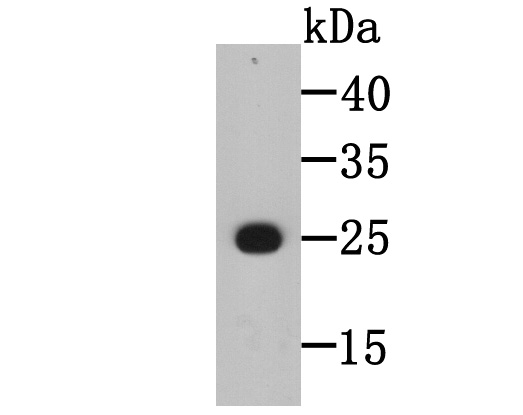
Fig1: Western blot analysis of BLCAP on recombinant protein tissue lysate using anti-BLCAP antibody at 1/5,000 dilution.Application
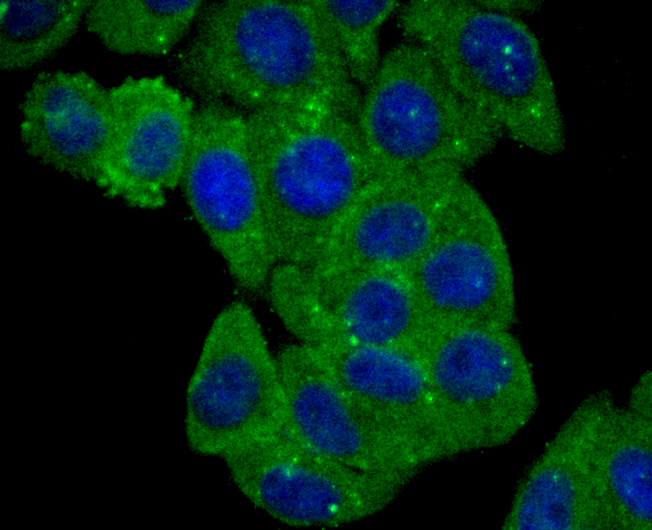
Fig2: ICC staining BLCAP (green) in HepG2 cells. The nuclear counter stain is DAPI (blue). Cells were fixed in paraformaldehyde, permeabilised with 0.25% Triton X100/PBS.Application
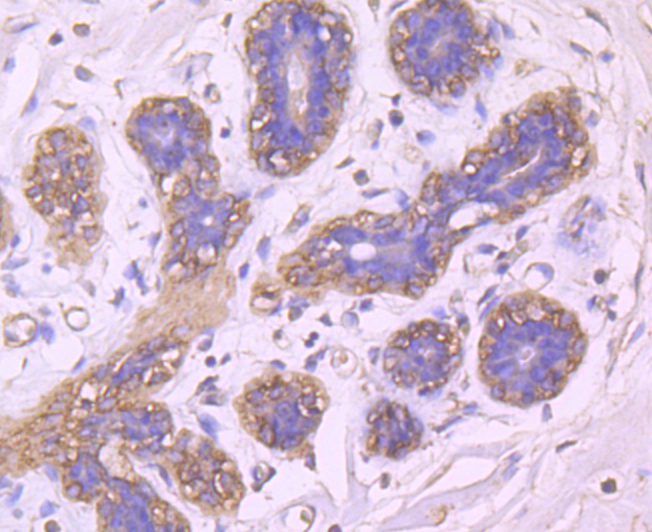
Fig3: Immunohistochemical analysis of paraffin-embedded human breast tissue using anti-BLCAP antibody. Counter stained with hematoxylin.Application
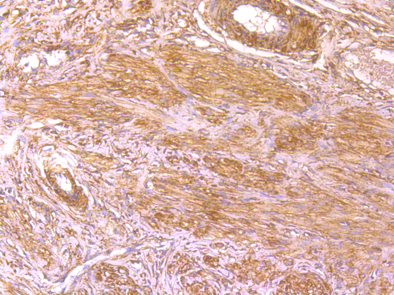
Fig4: Immunohistochemical analysis of paraffin-embedded rat cervix tissue using anti-BLCAP antibody. Counter stained with hematoxylin.| Product Name | BLCAP |
|---|---|
| Antibody Type | Primary Antibodies |
| Antigen Alias | Bc10 antibody Bladder cancer 10 kDa protein antibody Bladder cancer related protein (10kD) antibody Bladder cancer-associated protein antibody BLCAP antibody BLCAP_HUMAN antibody |
| Product description | BLCAP is a highly conversed gene with two exons and an intron encoding a 10kDa protein, which was originally identified from invasive bladder carcinoma in 2002. BLCAP is subject to adenosine to inosine (A-to-I) RNA editing. A-to-I RNA editing is an important post-transcription modification of RNA sequence, generating a diversity of RNA products different from the original DNA sequence. The adenosine deaminase acting on double-stranded RNA (ADAR) family of enzymes catalyze the conversion of adenosine (A) to inosine (I) in the double stranded RNA, and inosine is finally recognized as guanosine (G) in the process of mRNA translation. May regulate cell proliferation and coordinate apoptosis and cell cycle progression via a novel mechanism independent of both p53/TP53 and NF-kappa-B. |
| Immunogen | Peptide |
| Clonality | Monoclonal |
|---|---|
| Isotype | IgG2a |
| Host Species | Mouse |
| Tested Applications | WBIHCICC |
| WB:1:1,000-1:2,000 ICC:1:50-1:200 IHC:1:50-1:200 |
|
| Species Reactivity | HumanRat |
| Concentration | 2mg/ml |
| Alternative Names | Bc10 antibody Bladder cancer 10 kDa protein antibody Bladder cancer related protein (10kD) antibody Bladder cancer-associated protein antibody BLCAP antibody BLCAP_HUMAN antibody |
|---|---|
| Cellular Localization | Membrane. |
Application
Fig1: Western blot analysis of BLCAP on recombinant protein tissue lysate using anti-BLCAP antibody at 1/5,000 dilution.
Application
Fig2: ICC staining BLCAP (green) in HepG2 cells. The nuclear counter stain is DAPI (blue). Cells were fixed in paraformaldehyde, permeabilised with 0.25% Triton X100/PBS.
Application
Fig3: Immunohistochemical analysis of paraffin-embedded human breast tissue using anti-BLCAP antibody. Counter stained with hematoxylin.
Application
Fig4: Immunohistochemical analysis of paraffin-embedded rat cervix tissue using anti-BLCAP antibody. Counter stained with hematoxylin.| Positive Control | HepG2, rat cervix tissue, human breast tissue. |
|---|---|
| Application Notes | WB:1:1,000-1:2,000 ICC:1:50-1:200 IHC:1:50-1:200 |
| Form | Liquid |
|---|---|
| Storage Instructions | Store at +4℃ after thawing. Aliquot store at -20℃ or -80℃. Avoid repeated freeze / thaw cycles. |
| Storage Buffer | 1*TBS (pH7.4), 0.5%BSA, 50%Glycerol. Preservative: 0.05% Sodium Azide. |


