Application
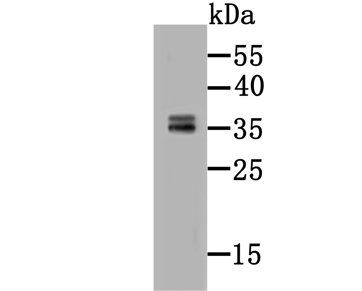
Fig1: Western blot analysis of CD137 on HepG2 cell lysate using anti-CD137 antibody at 1/500 dilution.Application
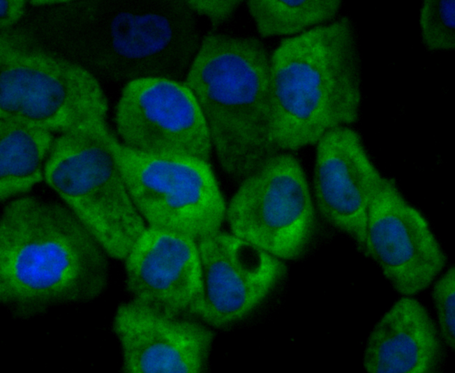
Fig2: ICC staining CD137 (green) in A431 cells. The nuclear counter stain is DAPI (blue). Cells were fixed in paraformaldehyde, permeabilised with 0.25% Triton X100/PBS.Application
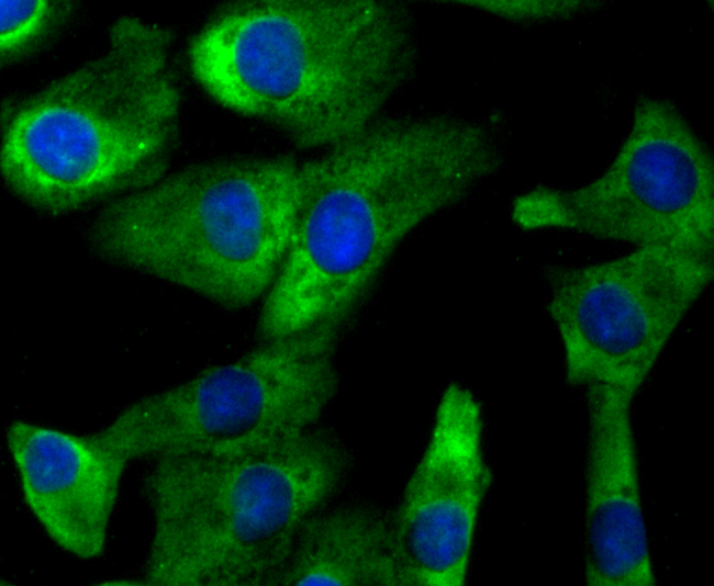
Fig3: ICC staining CD137 (green) in A549 cells. The nuclear counter stain is DAPI (blue). Cells were fixed in paraformaldehyde, permeabilised with 0.25% Triton X100/PBS.Application
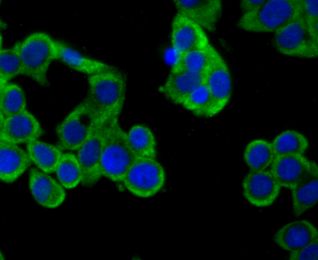
Fig4: ICC staining CD137 (green) in LOVO cells. The nuclear counter stain is DAPI (blue). Cells were fixed in paraformaldehyde, permeabilised with 0.25% Triton X100/PBS.Application
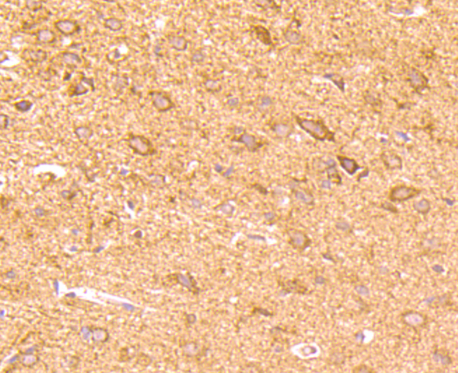
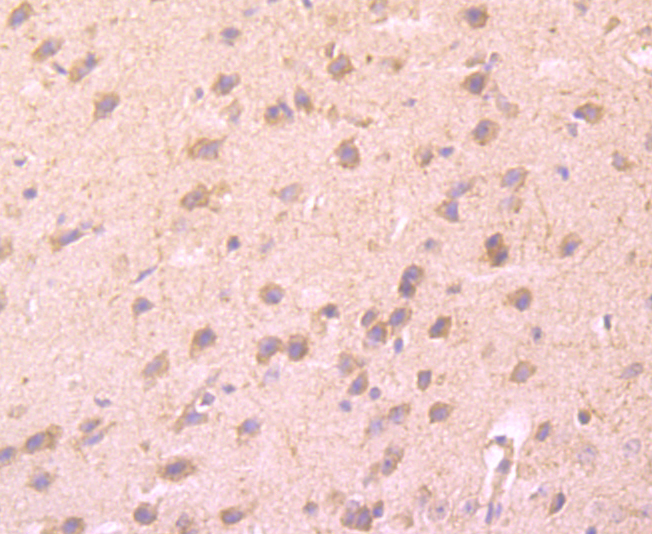
Fig5: Immunohistochemical analysis of paraffin-embedded rat brain tissue using anti-CD137 antibody. Counter stained with hematoxylin.Application
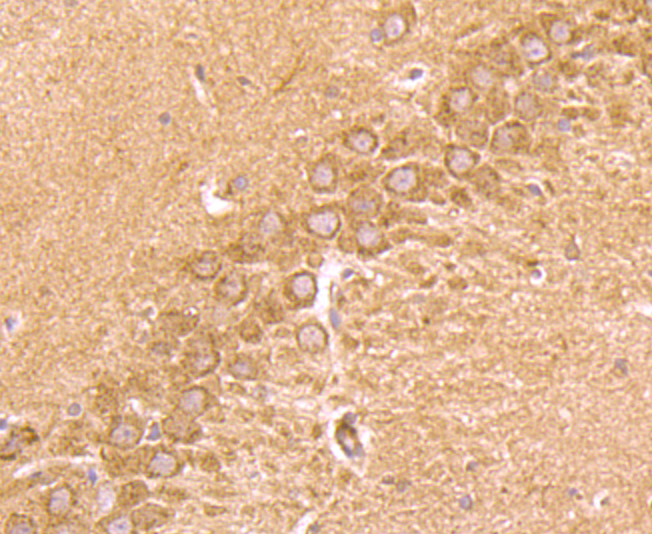
Fig6: Immunohistochemical analysis of paraffin-embedded rat hippocampus tissue using anti-CD137 antibody. Counter stained with hematoxylin.Application
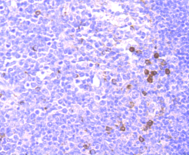
Fig7: Immunohistochemical analysis of paraffin-embedded human tonsil tissue using anti-CD137 antibody. Counter stained with hematoxylin.Application
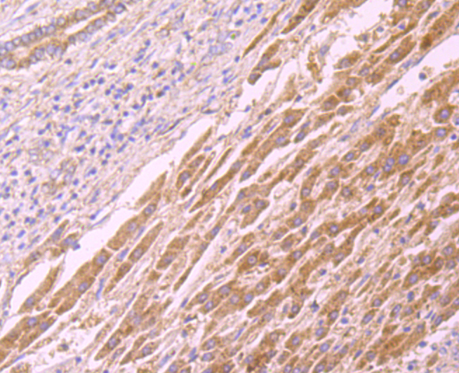
Fig8: Immunohistochemical analysis of paraffin-embedded human liver cancer tissue using anti-CD137 antibody. Counter stained with hematoxylin.Application
Fig9: Immunohistochemical analysis of paraffin-embedded mouse brain tissue using anti-CD137 antibody. Counter stained with hematoxylin.Application
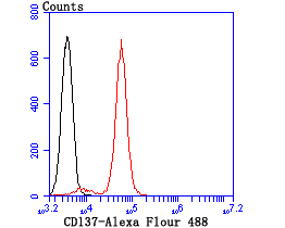
Fig10: Flow cytometric analysis of Jurkat cells with CD137 antibody at 1/100 dilution (red) compared with an unlabelled control (cells without incubation with primary antibody; black).| Product Name | CD137 [2G1] |
|---|---|
| Antibody Type | Primary Antibodies |
| Antigen Alias | 4 1BB antibody 4 1BB ligand receptor antibody 4-1BB ligand receptor antibody 4-1BB Ligand Receptor T Cell antibody 4-1BB, mouse, homolog of antibody Antigen 4-1BB Homolog antibody CD 137 antibody CD137 antibody CD137 antigen antibody CDw137 antibody HLDA VI antibody Homolog of mouse 4 1BB antibody ILA antibody induced by lymphocyte activation (ILA) antibody Induced by lymphocyte activation antibody Interleukin activated receptor homolog of mouse Ly63 antibody Ly63, mouse, homolog of antibody MGC2172 antibody OTTHUMP00000044294 antibody Receptor protein 4 1BB antibody T cell antigen 4 1BB homolog antibody T cell antigen ILA antibody T-cell antigen 4-1BB homolog antibody T-cell antigen ILA antibody TNF receptor superfamily member 9 antibody TNFRSF9 antibody TNR9_HUMAN antibody Tumor necrosis factor receptor superfamily member 9 antibody |
| Product description | CD137, also designated ILA and 4-1BB in mouse, belongs to the tumor necrosis factor receptor family and delivers a costimulatory signal to T lymphocytes. CD137 is expressed on activated T cells and binds an inducible ligand that is found on B cells, macrophages, and dendritic cells. Interactions between CD137 and its ligand are involved in antigen presentation and the generation of cytotoxic T cells. Crosslinking of the CD137 ligand induces apoptosis in resting lymphocytes. In contrast, CD137 regulates peripheral monocyte survival by inducing a cytokine release profile, and is mediated by M-CSF and to a lesser extent by granulocyte-macrophage colony-stimulating factor and IL-3. Soluble forms of CD137 are found in sera from patients with rheumatoid arthritis and may provide a negative control mechanism for immune responses. |
| Immunogen | Recombinant protein |
| Clonality | Monoclonal |
|---|---|
| Isotype | IgG1 |
| Host Species | Mouse |
| Tested Applications | WBIHCICCFC |
| WB:1:500 ICC:1:100-1:500 IHC:1:50-1:200 FC:1:50-1:200 |
|
| Species Reactivity | HumanMouseRat |
| Concentration | 2mg/ml |
| Alternative Names | 4 1BB antibody
4 1BB ligand receptor antibody
4-1BB ligand receptor antibody
4-1BB Ligand Receptor T Cell antibody
4-1BB mouse homolog of antibody Antigen 4-1BB Homolog antibody CD 137 antibody CD137 antibody CD137 antigen antibody CDw137 antibody HLDA VI antibody Homolog of mouse 4 1BB antibody ILA antibody induced by lymphocyte activation (ILA) antibody Induced by lymphocyte activation antibody Interleukin activated receptor homolog of mouse Ly63 antibody Ly63 mouse homolog of antibody MGC2172 antibody OTTHUMP00000044294 antibody Receptor protein 4 1BB antibody T cell antigen 4 1BB homolog antibody T cell antigen ILA antibody T-cell antigen 4-1BB homolog antibody T-cell antigen ILA antibody TNF receptor superfamily member 9 antibody TNFRSF9 antibody TNR9_HUMAN antibody Tumor necrosis factor receptor superfamily member 9 antibody |
|---|---|
| Molecular Weight(MW) | 35 kDa |
| Cellular Localization | Membrane. Extracellular space. |
Application
Fig1: Western blot analysis of CD137 on HepG2 cell lysate using anti-CD137 antibody at 1/500 dilution.
Application
Fig2: ICC staining CD137 (green) in A431 cells. The nuclear counter stain is DAPI (blue). Cells were fixed in paraformaldehyde, permeabilised with 0.25% Triton X100/PBS.
Application
Fig3: ICC staining CD137 (green) in A549 cells. The nuclear counter stain is DAPI (blue). Cells were fixed in paraformaldehyde, permeabilised with 0.25% Triton X100/PBS.
Application
Fig4: ICC staining CD137 (green) in LOVO cells. The nuclear counter stain is DAPI (blue). Cells were fixed in paraformaldehyde, permeabilised with 0.25% Triton X100/PBS.
Application
Fig5: Immunohistochemical analysis of paraffin-embedded rat brain tissue using anti-CD137 antibody. Counter stained with hematoxylin.
Application
Fig6: Immunohistochemical analysis of paraffin-embedded rat hippocampus tissue using anti-CD137 antibody. Counter stained with hematoxylin.
Application
Fig7: Immunohistochemical analysis of paraffin-embedded human tonsil tissue using anti-CD137 antibody. Counter stained with hematoxylin.
Application
Fig8: Immunohistochemical analysis of paraffin-embedded human liver cancer tissue using anti-CD137 antibody. Counter stained with hematoxylin.
Application
Fig9: Immunohistochemical analysis of paraffin-embedded mouse brain tissue using anti-CD137 antibody. Counter stained with hematoxylin.
Application
Fig10: Flow cytometric analysis of Jurkat cells with CD137 antibody at 1/100 dilution (red) compared with an unlabelled control (cells without incubation with primary antibody; black).| Positive Control | A431, A549, LOVO, rat brain tissue, rat hippocampus tissue, human liver cancer tissue, mouse brain tissue, human tonsil tissue, Jurkat. |
|---|---|
| Application Notes | WB:1:500 ICC:1:100-1:500 IHC:1:50-1:200 FC:1:50-1:200 |
| Form | Liquid |
|---|---|
| Storage Instructions | Store at +4℃ after thawing. Aliquot store at -20℃ or -80℃. Avoid repeated freeze / thaw cycles. |
| Storage Buffer | 1*TBS (pH7.4), 0.5%BSA, 50%Glycerol. Preservative: 0.05% Sodium Azide. |


