Application
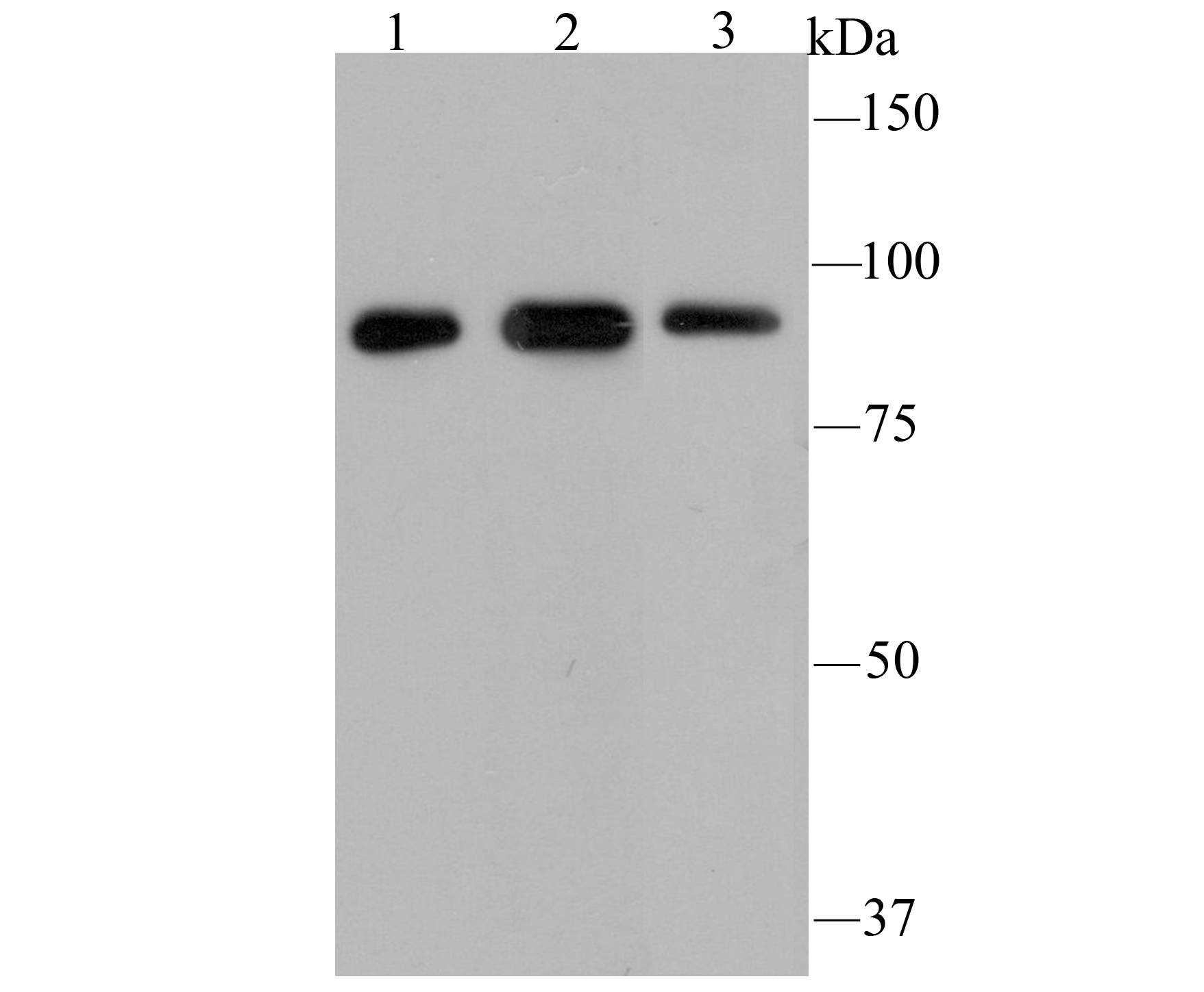
Fig1: Western blot analysis of GRP94 on different cell lysates using anti-GRP94 antibody at 1/5,000 dilution. Positive control: Lane1: NIH-3T3 Lane2: Hela Lane3: HepG2Application
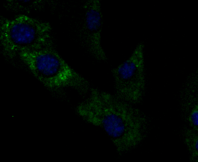
Fig2: ICC staining GRP94 (green) in NIH-3T3 cells. The nuclear counter stain is DAPI (blue). Cells were fixed in paraformaldehyde, permeabilised with 0.25% Triton X100/PBS.Application
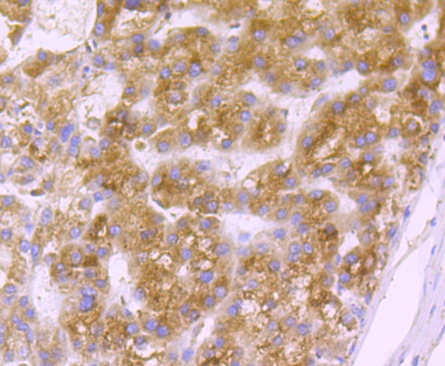
Fig3: Immunohistochemical analysis of paraffin-embedded human liver cancer tissue using anti-GRP94 antibody. Counter stained with hematoxylin.Application
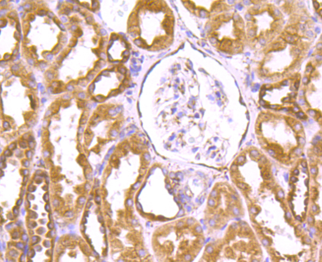
Fig4: Immunohistochemical analysis of paraffin-embedded human kidney tissue using anti-GRP94 antibody. Counter stained with hematoxylin.Application
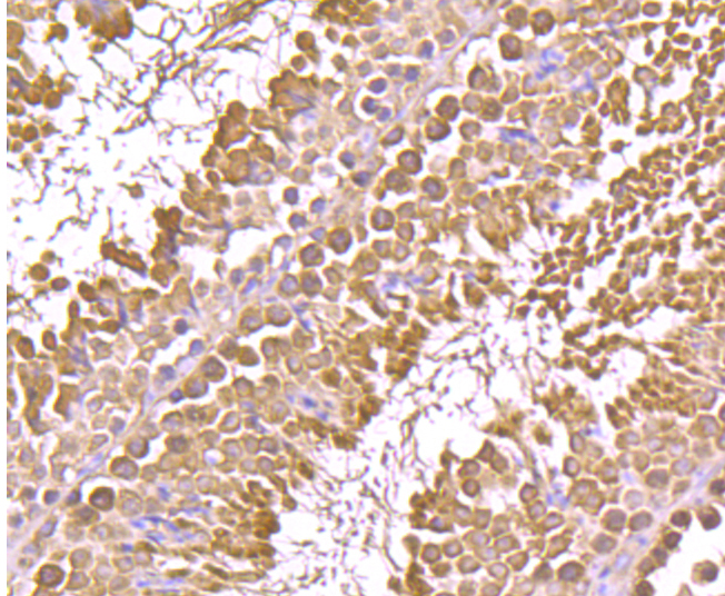
Fig5: Immunohistochemical analysis of paraffin-embedded mouse testis tissue using anti-GRP94 antibody. Counter stained with hematoxylin.Application
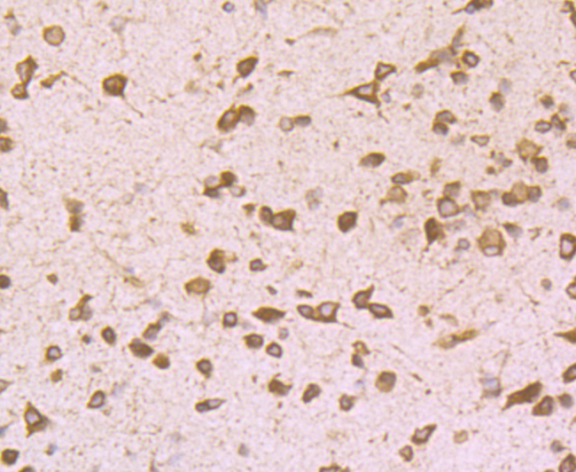
Fig6: Immunohistochemical analysis of paraffin-embedded mouse brain tissue using anti-GRP94 antibody. Counter stained with hematoxylin.| Product Name | GRP94 [F1-D10-G3] |
|---|---|
| Antibody Type | Primary Antibodies |
| Antigen Alias | 94 kDa glucose regulated protein antibody 94 kDa glucose-regulated protein antibody ECGP antibody Endoplasmin antibody Endothelial cell (HBMEC) glycoprotein antibody ENPL_HUMAN antibody Glucose regulated protein 94kDa antibody gp96 antibody gp96 homolog antibody GRP 94 antibody GRP-94 antibody Heat shock protein 90 kDa beta member 1 antibody heat shock protein 90kDa beta (Grp94), member 1 antibody Heat shock protein, 90 kDa, beta, 1 antibody HSP90B1 antibody Stress inducible tumor rejection antigen GP96 antibody TRA1 antibody tumor rejection antigen (gp96) 1 antibody Tumor rejection antigen 1 antibody Tumor rejection antigen gp96 antibody Tumor rejection antigen-1 (gp96) antibody |
| Product description | The human HSP 90 family includes 17 genes that fall into four classes: HSP90AA, HSP90AB, HSP90B and TRAP. HSP 90 family members guide the normal folding, intracellular disposition and Proteolytic turnover of many key regulators of cell growth, differentiation and survival. HSP 90α, also designated HSP90A, HSP 86 and LPS-associated protein 2 (LAP2), is a cytosolic enhancer of inducible nitric-oxide synthase (iNOS), with chaperone activity that is important for the transcriptional activity of p53. HSP 90β, also designated HSP90B, HSP 84 and HSPC2, is a cytosolic protein that participates in signaling pathways with PKC e to protect cells from external damage, particularly in heat shock-mediated events. GRP 94, also known as tumor rejection antigen 1 (TRA1), ECGP and GP96, localizes to the ER, is highly expressed in BGC-823 human gastric carcinoma cells and is upregulated in human endothelial cells in response to hypoxia by HIF-1. |
| Immunogen | Recombinant protein |
| Clonality | Monoclonal |
|---|---|
| Isotype | IgM |
| Host Species | Mouse |
| Tested Applications | WBIHCICC |
| WB:1:1,000-1:10,000 ICC:1:50 IHC:1:50-1:200 |
|
| Species Reactivity | HumanMouse |
| Concentration | 2mg/ml |
| Alternative Names | 94 kDa glucose regulated protein antibody
94 kDa glucose-regulated protein antibody
ECGP antibody
Endoplasmin antibody
Endothelial cell (HBMEC) glycoprotein antibody
ENPL_HUMAN antibody
Glucose regulated protein 94kDa antibody
gp96 antibody
gp96 homolog antibody
GRP 94 antibody
GRP-94 antibody
Heat shock protein 90 kDa beta member 1 antibody
heat shock protein 90kDa beta (Grp94) member 1 antibody Heat shock protein 90 kDa beta 1 antibody HSP90B1 antibody Stress inducible tumor rejection antigen GP96 antibody TRA1 antibody tumor rejection antigen (gp96) 1 antibody Tumor rejection antigen 1 antibody Tumor rejection antigen gp96 antibody Tumor rejection antigen-1 (gp96) antibody |
|---|---|
| Molecular Weight(MW) | 94 kDa |
| Cellular Localization | Endoplasmic reticulum lumen, melanosome. |
Application
Fig1: Western blot analysis of GRP94 on different cell lysates using anti-GRP94 antibody at 1/5,000 dilution. Positive control: Lane1: NIH-3T3 Lane2: Hela Lane3: HepG2
Application
Fig2: ICC staining GRP94 (green) in NIH-3T3 cells. The nuclear counter stain is DAPI (blue). Cells were fixed in paraformaldehyde, permeabilised with 0.25% Triton X100/PBS.
Application
Fig3: Immunohistochemical analysis of paraffin-embedded human liver cancer tissue using anti-GRP94 antibody. Counter stained with hematoxylin.
Application
Fig4: Immunohistochemical analysis of paraffin-embedded human kidney tissue using anti-GRP94 antibody. Counter stained with hematoxylin.
Application
Fig5: Immunohistochemical analysis of paraffin-embedded mouse testis tissue using anti-GRP94 antibody. Counter stained with hematoxylin.
Application
Fig6: Immunohistochemical analysis of paraffin-embedded mouse brain tissue using anti-GRP94 antibody. Counter stained with hematoxylin.| Positive Control | HepG2, Hela, NIH-3T3, human liver cancer tissue, human kidney tissue, mouse testis tissue, mouse brain tissue. |
|---|---|
| Application Notes | WB:1:1,000-1:10,000 ICC:1:50 IHC:1:50-1:200 |
| Form | Liquid |
|---|---|
| Storage Instructions | Store at +4℃ after thawing. Aliquot store at -20℃ or -80℃. Avoid repeated freeze / thaw cycles. |
| Storage Buffer | 1*TBS (pH7.4), 0.5%BSA, 50%Glycerol. Preservative: 0.05% Sodium Azide. |


