Application
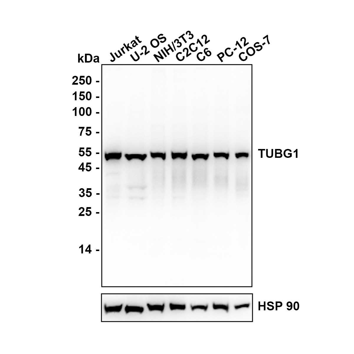
Fig1: Western blot analysis of gamma tubulin on different lysates with Rabbit anti-gamma tubulin antibody at 1/2,000 dilution. Lane 1: Jurkat, Lane 2: U-2 OS, Lane 3: NIH/3T3, Lane 4: C2C12, Lane 5: C6, Lane 6: PC-12, Lane 7: COS-7, Lysates/proteins at 20 µg/Lane. Exposure time: 15 seconds; Blocking: 5% NFDM/TBST, 1 hour at room temperature Primary antibody: 1/2,000 in primary antibody dilution buffer, overnight at 4 ℃ Secondary antibody: Goat anti-Mouse IgG-HRP, 1/50,000 in 5% NFDM/TBST, 1 hour at room temperature.Application
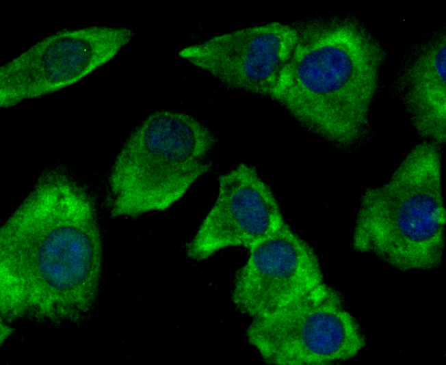
Fig2: ICC staining gamma tubulin (green) in A549 cells. The nuclear counter stain is DAPI (blue). Cells were fixed in paraformaldehyde, permeabilised with 0.25% Triton X100/PBS.Application
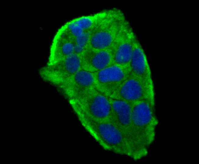
Fig3: ICC staining gamma tubulin (green) in Hela cells. The nuclear counter stain is DAPI (blue). Cells were fixed in paraformaldehyde, permeabilised with 0.25% Triton X100/PBS.Application
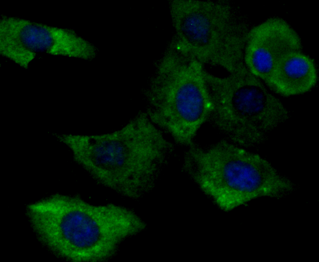
Fig4: ICC staining gamma tubulin (green) in NIH-3T3 cells. The nuclear counter stain is DAPI (blue). Cells were fixed in paraformaldehyde, permeabilised with 0.25% Triton X100/PBS.Application
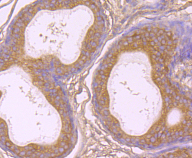
Fig5: Immunohistochemical analysis of paraffin-embedded rat epididymis tissue using anti-gamma tubulin antibody. Counter stained with hematoxylin.Application
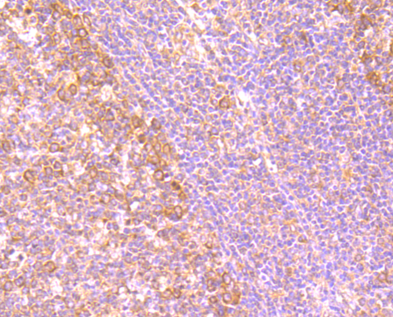
Fig6: Immunohistochemical analysis of paraffin-embedded human tonsil tissue using anti-gamma tubulin antibody. Counter stained with hematoxylin.Application
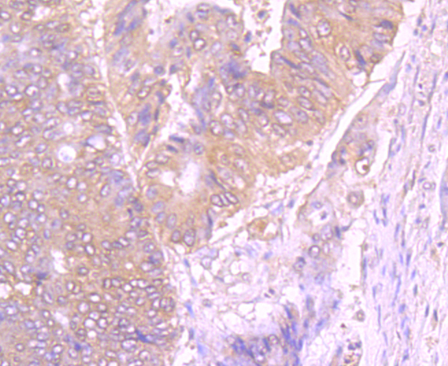
Fig7: Immunohistochemical analysis of paraffin-embedded human colon cancer tissue using anti-gamma tubulin antibody. Counter stained with hematoxylin.Application
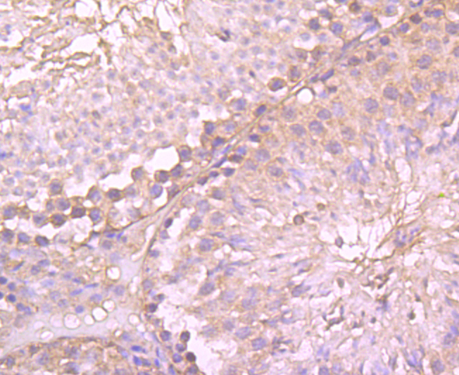
Fig8: Immunohistochemical analysis of paraffin-embedded mouse testis tissue using anti-gamma tubulin antibody. Counter stained with hematoxylin.| Product Name | gamma tubulin Mouse Monoclonal Antibody |
|---|---|
| Antibody Type | Primary Antibodies |
| Product description | Tubulin is a major cytoskeleton component that has five distinct forms, designated α, β, γ, δ and e Tubulin. α and β Tubulins form heterodimers which multimerize to form a microtubule filament. Multiple β Tubulin isoforms (β1, β2, β3, β4, β5, β6 and β8) have been characterized and are expressed in mammalian tissues. β1 and β4 are present throughout the cytosol, β2 is present in the nuclei and nucleoplasm, and β3 is a neuron-specific cytoskeletal protein. γ Tubulin forms the gammasome, which is required for nucleating microtubule filaments at the centrosome. Both δ Tubulin and e Tubulin are associated with the centrosome. δ Tubulin is a homolog of the Chlamydomonas δ Tubulin Uni3 and is found in association with the centrioles, whereas e Tubulin localizes to the pericentriolar material. e Tubulin exhibits a cell-cycle-specific pattern of localization, first associating with only the older of the centrosomes in a newly duplicated pair and later associating with both centrosomes. |
| Immunogen | Peptide |
| Clonality | Monoclonal |
|---|---|
| Isotype | IgG2a |
| Host Species | Mouse |
| Tested Applications | ICCIHCWB |
| WB:1:1,000-1:5,000 ICC:1:100-1:500 IHC:1:50-1:200 |
|
| Species Reactivity | HumanMouseRat |
| Concentration | 1mg/ml |
| Purification | Protein G |
| Alternative Names | Gamma 1 tubulin antibody
Gamma 2 tubulin antibody
Gamma Tubulin 1 antibody
Gamma Tubulin 2 antibody
Gamma tubulin complex component 1 antibody
Gamma-2-tubulin antibody
GCP 1 antibody
GCP1 antibody
MGC131994 antibody
TBG2_HUMAN antibody
TUBG antibody
TUBG1 antibody
TUBG2 antibody
TUBGCP1 antibody
Tubulin gamma 1 chain antibody
Tubulin gamma 2 chain antibody
Tubulin gamma complex-associated protein 1 antibody
Tubulin gamma-2 chain antibody
tubulin gamma 1 antibody tubulin gamma 2 antibody tubulin gamma polypeptide antibody Xgam antibody |
|---|---|
| Molecular Weight(MW) | 51 kDa |
| Cellular Localization | Cytoplasm, cytoskeleton, centrosome. |
Application
Fig1: Western blot analysis of gamma tubulin on different lysates with Rabbit anti-gamma tubulin antibody at 1/2,000 dilution. Lane 1: Jurkat, Lane 2: U-2 OS, Lane 3: NIH/3T3, Lane 4: C2C12, Lane 5: C6, Lane 6: PC-12, Lane 7: COS-7, Lysates/proteins at 20 µg/Lane. Exposure time: 15 seconds; Blocking: 5% NFDM/TBST, 1 hour at room temperature Primary antibody: 1/2,000 in primary antibody dilution buffer, overnight at 4 ℃ Secondary antibody: Goat anti-Mouse IgG-HRP, 1/50,000 in 5% NFDM/TBST, 1 hour at room temperature.
Application
Fig2: ICC staining gamma tubulin (green) in A549 cells. The nuclear counter stain is DAPI (blue). Cells were fixed in paraformaldehyde, permeabilised with 0.25% Triton X100/PBS.
Application
Fig3: ICC staining gamma tubulin (green) in Hela cells. The nuclear counter stain is DAPI (blue). Cells were fixed in paraformaldehyde, permeabilised with 0.25% Triton X100/PBS.
Application
Fig4: ICC staining gamma tubulin (green) in NIH-3T3 cells. The nuclear counter stain is DAPI (blue). Cells were fixed in paraformaldehyde, permeabilised with 0.25% Triton X100/PBS.
Application
Fig5: Immunohistochemical analysis of paraffin-embedded rat epididymis tissue using anti-gamma tubulin antibody. Counter stained with hematoxylin.
Application
Fig6: Immunohistochemical analysis of paraffin-embedded human tonsil tissue using anti-gamma tubulin antibody. Counter stained with hematoxylin.
Application
Fig7: Immunohistochemical analysis of paraffin-embedded human colon cancer tissue using anti-gamma tubulin antibody. Counter stained with hematoxylin.
Application
Fig8: Immunohistochemical analysis of paraffin-embedded mouse testis tissue using anti-gamma tubulin antibody. Counter stained with hematoxylin.| Application Notes | WB:1:1,000-1:5,000 ICC:1:100-1:500 IHC:1:50-1:200 |
|---|
| Form | Liquid |
|---|---|
| Storage Instructions | Store at +4℃ after thawing. Aliquot store at -20℃ or -80℃. Avoid repeated freeze / thaw cycles. |
| Storage Buffer | 1*TBS (pH7.4), 0.5%BSA, 50%Glycerol. Preservative: 0.05% Sodium Azide. |


