Application
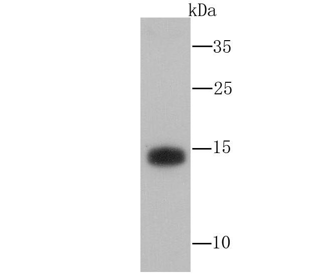
Fig1: Western blot analysis of Atg12 on THP-1 cell lysate using anti-Atg12 antibody atApplication
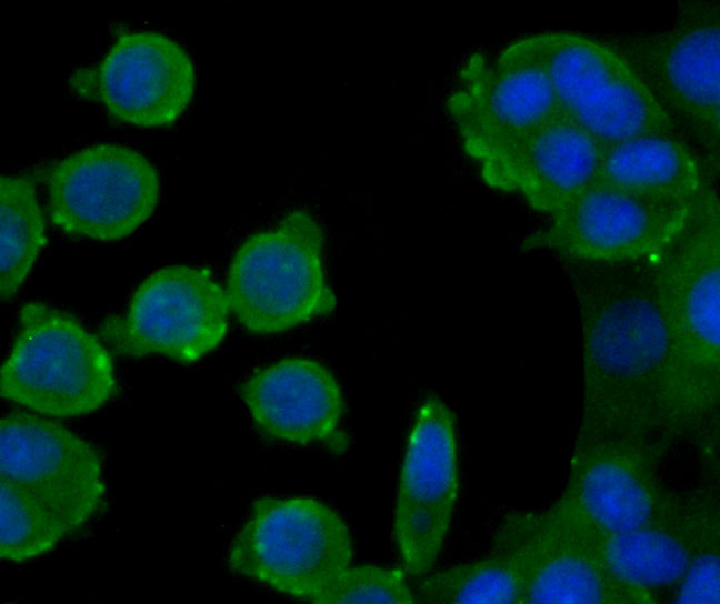
Fig2: ICC staining Atg12 (green) in MCF-7 cells. The nuclear counter stain is DAPI (blue). Cells were fixed in paraformaldehyde, permeabilised with 0.25% Triton X100/PBS.Application
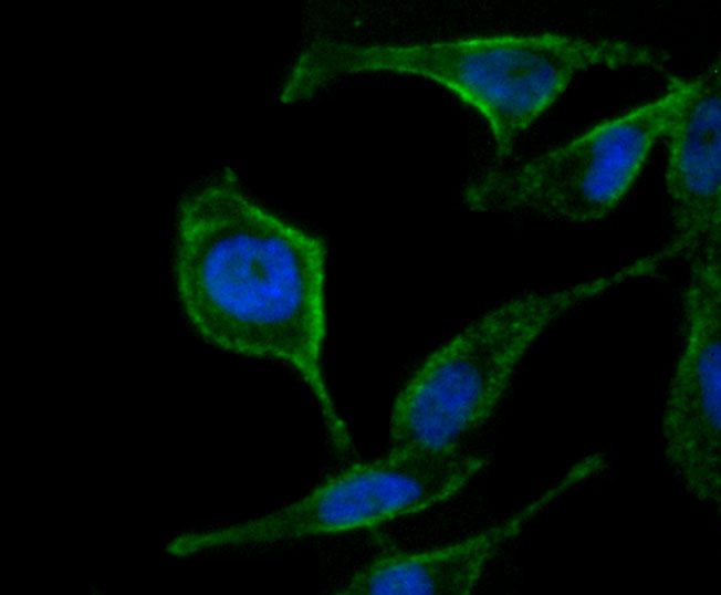
Fig3: ICC staining Atg12 (green) in PC-3M cells. The nuclear counter stain is DAPI (blue). Cells were fixed in paraformaldehyde, permeabilised with 0.25% Triton X100/PBS.Application
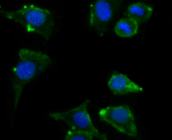
Fig4: ICC staining Atg12 (green) in SH-SY5Y cells. The nuclear counter stain is DAPI (blue). Cells were fixed in paraformaldehyde, permeabilised with 0.25% Triton X100/PBS.Application
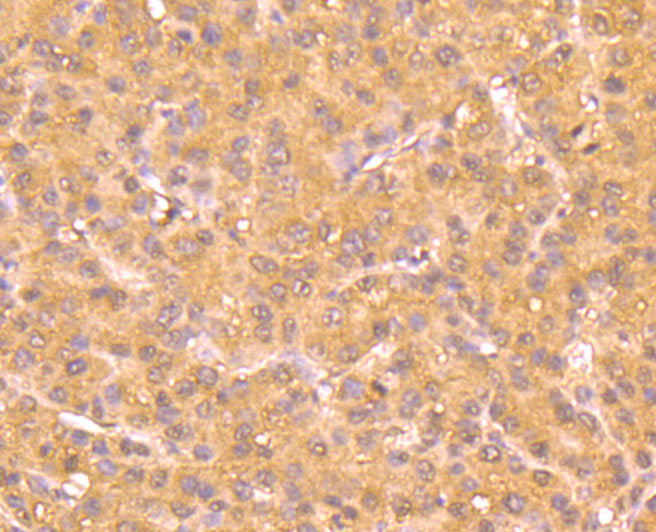
Fig5: Immunohistochemical analysis of paraffin-embedded human liver cancer tissue using anti-Atg12 antibody. Counter stained with hematoxylin.Application
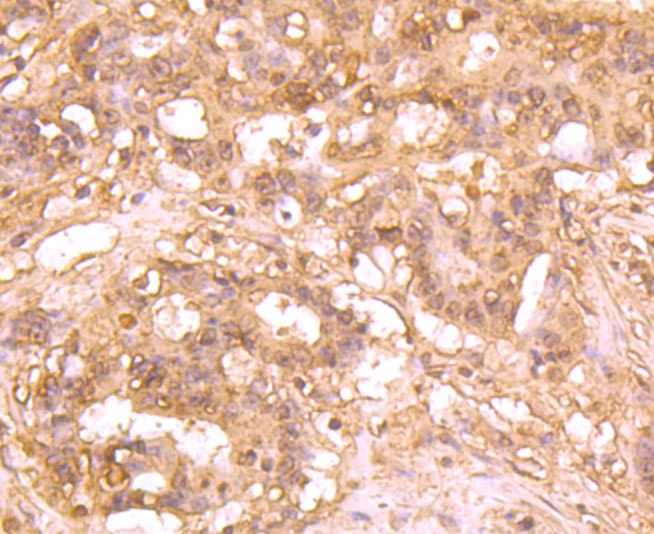
Fig6: Immunohistochemical analysis of paraffin-embedded human stomach cancer tissue using anti-Atg12 antibody. Counter stained with hematoxylin.Application
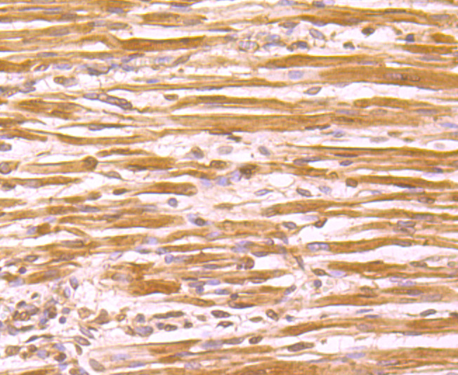
Fig7: Immunohistochemical analysis of paraffin-embedded human fetal skeletal muscle tissue using anti-Atg12 antibody. Counter stained with hematoxylin.| Product Name | Atg12 [1-11] |
|---|---|
| Antibody Type | Primary Antibodies |
| Antigen Alias | APG12-like antibody APG12L antibody ATG12 antibody ATG12 autophagy related 12 homolog (S. cerevisiae) antibody ATG12 autophagy related 12 homolog antibody ATG12_HUMAN antibody Autophagy 12 antibody Autophagy-related protein 12 antibody FBR93 antibody HAPG12 antibody Ubiquitin-like protein ATG12 antibody |
| Product description | Atg12 (autophagy-related protein 12), also known as APG12, APG12L, FBR93 or HAPG12, is a 140 amino acid protein that is ubiquitously expressed and belongs to the Atg12 family of proteins. Atg12 is a homolog of the yeast protein Apg12 that participates in autophagy. Autophagy is a membrane trafficking mechanism that delivers cytoplasmic cargo to the vacuole/lysosome for degradation and recycling. In yeast, autophagy requires a protein conjugation system consisting of Apg12 covalently bound at the carboxy terminal glycine to lysine 149 of Apg5. Similarly in humans, Atg12 is essential for autophagy and localizes to the cytoplasm where it is covalently bound to APG5, a conjugation reaction that requires APG7, Atg10 and ATP. The Atg12-APG5 conjugate functions as an important regulator of the autophagic process and is required for the change in membrane morphology and development of autophagosomes. Due to alternative splicing events, two Atg12 isoforms exist. |
| Immunogen | Peptide |
| Clonality | Monoclonal |
|---|---|
| Isotype | IgE |
| Host Species | Mouse |
| Tested Applications | WBIHCICC |
| WB:1:500-1:1,000 ICC:1:50-1:200 IHC:1:50-1:200 |
|
| Species Reactivity | HumanMouseRat |
| Concentration | 2mg/ml |
| Alternative Names | APG12-like antibody APG12L antibody ATG12 antibody ATG12 autophagy related 12 homolog (S. cerevisiae) antibody ATG12 autophagy related 12 homolog antibody ATG12_HUMAN antibody Autophagy 12 antibody Autophagy-related protein 12 antibody FBR93 antibody HAPG12 antibody Ubiquitin-like protein ATG12 antibody |
|---|---|
| Molecular Weight(MW) | 15 kDa |
| Cellular Localization | Cytoplasm. Membrane. |
Application
Fig1: Western blot analysis of Atg12 on THP-1 cell lysate using anti-Atg12 antibody at
Application
Fig2: ICC staining Atg12 (green) in MCF-7 cells. The nuclear counter stain is DAPI (blue). Cells were fixed in paraformaldehyde, permeabilised with 0.25% Triton X100/PBS.
Application
Fig3: ICC staining Atg12 (green) in PC-3M cells. The nuclear counter stain is DAPI (blue). Cells were fixed in paraformaldehyde, permeabilised with 0.25% Triton X100/PBS.
Application
Fig4: ICC staining Atg12 (green) in SH-SY5Y cells. The nuclear counter stain is DAPI (blue). Cells were fixed in paraformaldehyde, permeabilised with 0.25% Triton X100/PBS.
Application
Fig5: Immunohistochemical analysis of paraffin-embedded human liver cancer tissue using anti-Atg12 antibody. Counter stained with hematoxylin.
Application
Fig6: Immunohistochemical analysis of paraffin-embedded human stomach cancer tissue using anti-Atg12 antibody. Counter stained with hematoxylin.
Application
Fig7: Immunohistochemical analysis of paraffin-embedded human fetal skeletal muscle tissue using anti-Atg12 antibody. Counter stained with hematoxylin.| Positive Control | THP-1, MCF-7. PC-3M, SH-SY5Y, human liver cancer tissue, human stomach cancer tissue, human fetal skeletal muscle tissue. |
|---|---|
| Application Notes | WB:1:500-1:1,000 ICC:1:50-1:200 IHC:1:50-1:200 |
| Form | Liquid |
|---|---|
| Storage Instructions | Store at +4℃ after thawing. Aliquot store at -20℃ or -80℃. Avoid repeated freeze / thaw cycles. |
| Storage Buffer | 1*TBS (pH7.4), 1%BSA, 40%Glycerol. Preservative: 0.05% Sodium Azide. |


