Application
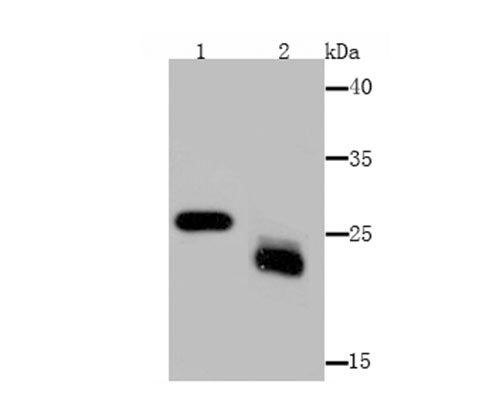
Fig1: Western blot analysis of MAL on mouse kidney (1) and mouse lymphatic vessels (2) tissue lysate using anti-MAL antibody at 1/1,000 dilution.Application
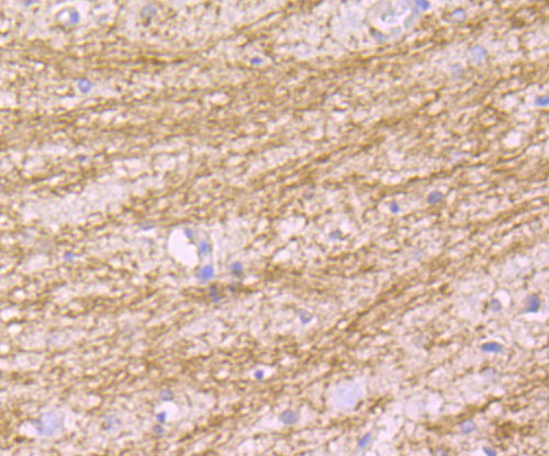
Fig2: Immunohistochemical analysis of paraffin-embedded rat brain tissue using anti-MAL antibody. Counter stained with hematoxylin.Application
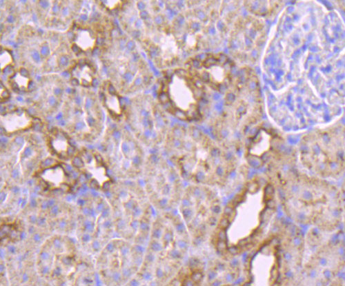
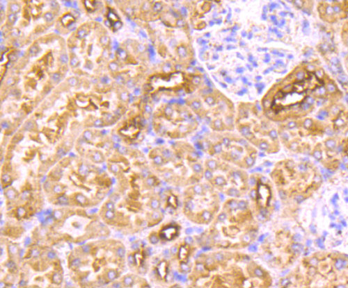
Fig3: Immunohistochemical analysis of paraffin-embedded rat kidney tissue using anti-MAL antibody. Counter stained with hematoxylin.Application
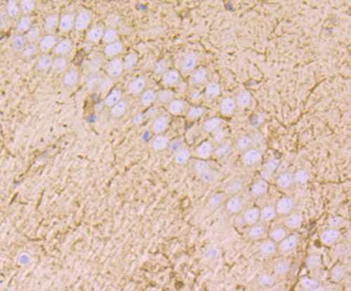
Fig4: Immunohistochemical analysis of paraffin-embedded mouse brain tissue using anti-MAL antibody. Counter stained with hematoxylin.Application
Fig5: Immunohistochemical analysis of paraffin-embedded mouse kidney tissue using anti-MAL antibody. Counter stained with hematoxylin.Application
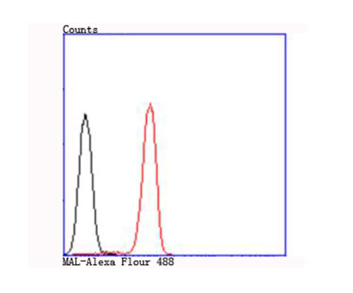
Fig6: Flow cytometric analysis of Jurkat cells with MAL antibody at 1/100 dilution (red) compared with an unlabelled control (cells without incubation with primary antibody; black).| Product Name | MAL [B5-G3] |
|---|---|
| Antibody Type | Primary Antibodies |
| Antigen Alias | mal antibody MAL protein gene antibody Mal T-cell differentiation protein antibody MAL_HUMAN antibody MALGENE antibody MPV17 antibody Myelin and lymphocyte protein antibody T-cell differentiation protein MAL antibody T-lymphocyte maturation-associated protein antibody VIP17 antibody |
| Product description | MAL (myelin and lymphocyte protein), also known as T lymphocyte maturation-associated protein, is a nonglycosylated hydrophobic integral membrane protein belonging to the MAL family of proteolipids. MAL is highly enriched in nervous system myelin and in rafts and apical membranes of epithelial cells. It is involved in forming, stabilizing and maintaining glycosphingolipid-enriched membrane microdomains. MAL maintains the myelin sheath and, by controlling the sorting and trafficking of oligodendrocytes, it is involved in central nervous system paranode maintenance. MAL is a component of lipid rafts in myelinating cells. Association with glycosphingolipids may result in protein-lipid microdomain formation in myelin. MAL has been localized to the endoplasmic reticulum of T cells and in compact myelin of cells in the nervous system. MAL is primarily expressed by oligodendrocytes and Schwann cells in the intermediate and late stages of T cell differentiation. |
| Immunogen | Peptide |
| Clonality | Monoclonal |
|---|---|
| Isotype | IgG1 |
| Host Species | Mouse |
| Tested Applications | WBIHCFC |
| WB:1:250~1:1000 IHC:1:20~1:100 FC:1:100~1:200 Notes:Optimal dilutions/concentrations should be determined by the researcher. |
|
| Species Reactivity | HumanMouseRat |
| Concentration | 2mg/ml |
| Alternative Names | mal antibody MAL protein gene antibody Mal T-cell differentiation protein antibody MAL_HUMAN antibody MALGENE antibody MPV17 antibody Myelin and lymphocyte protein antibody T-cell differentiation protein MAL antibody T-lymphocyte maturation-associated protein antibody VIP17 antibody |
|---|---|
| Molecular Weight(MW) | 25 kDa |
| Cellular Localization | Membrane. |
Application
Fig1: Western blot analysis of MAL on mouse kidney (1) and mouse lymphatic vessels (2) tissue lysate using anti-MAL antibody at 1/1,000 dilution.
Application
Fig2: Immunohistochemical analysis of paraffin-embedded rat brain tissue using anti-MAL antibody. Counter stained with hematoxylin.
Application
Fig3: Immunohistochemical analysis of paraffin-embedded rat kidney tissue using anti-MAL antibody. Counter stained with hematoxylin.
Application
Fig4: Immunohistochemical analysis of paraffin-embedded mouse brain tissue using anti-MAL antibody. Counter stained with hematoxylin.
Application
Fig5: Immunohistochemical analysis of paraffin-embedded mouse kidney tissue using anti-MAL antibody. Counter stained with hematoxylin.
Application
Fig6: Flow cytometric analysis of Jurkat cells with MAL antibody at 1/100 dilution (red) compared with an unlabelled control (cells without incubation with primary antibody; black).| Positive Control | Mouse kidney tissue, mouse lymphatic vessels tissue lysate, rat brain tissue, rat kidney tissue, mouse brain tissue, Jurkat. |
|---|---|
| Application Notes | WB:1:250~1:1000 IHC:1:20~1:100 FC:1:100~1:200 Notes:Optimal dilutions/concentrations should be determined by the researcher. |
| Form | Liquid |
|---|---|
| Storage Instructions | Store at +4℃ after thawing. Aliquot store at -20℃ or -80℃. Avoid repeated freeze / thaw cycles. |
| Storage Buffer | 1*TBS (pH7.4), 0.5%BSA, 50%Glycerol. Preservative: 0.05% Sodium Azide. |


