Application
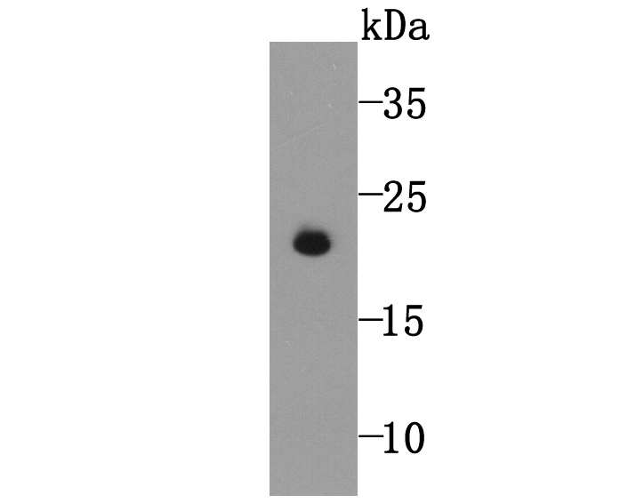
Fig1: Western blot analysis of GPX5 on human sperm tissue lysate using anti-GPX5 antibody at 1/5,000 dilution.Application
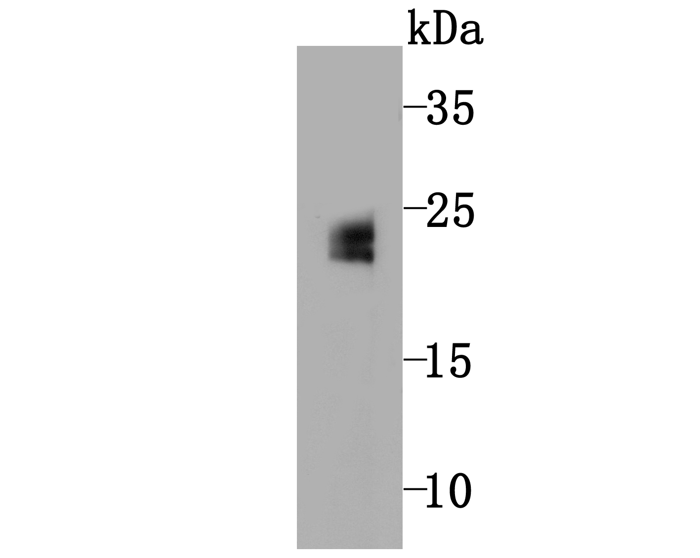
Fig2: Western blot analysis of GPX5 on mouse testis tissue lysate using anti-GPX5 antibody at 1/500 dilution.Application
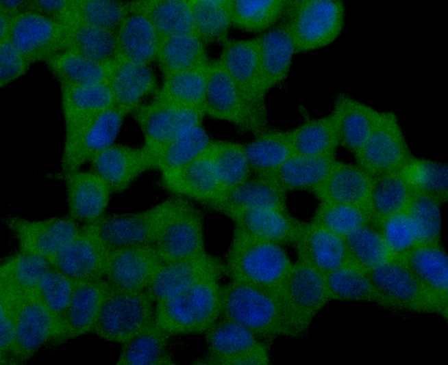
Fig3: ICC staining GPX5 (green) in 293T cells. The nuclear counter stain is DAPI (blue). Cells were fixed in paraformaldehyde, permeabilised with 0.25% Triton X100/PBS.Application
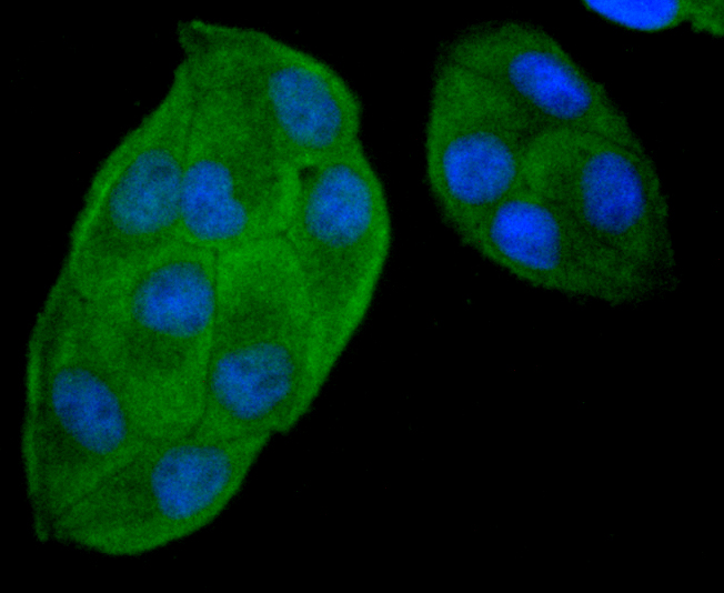
Fig4: ICC staining GPX5 (green) in Hela cells. The nuclear counter stain is DAPI (blue). Cells were fixed in paraformaldehyde, permeabilised with 0.25% Triton X100/PBS.Application
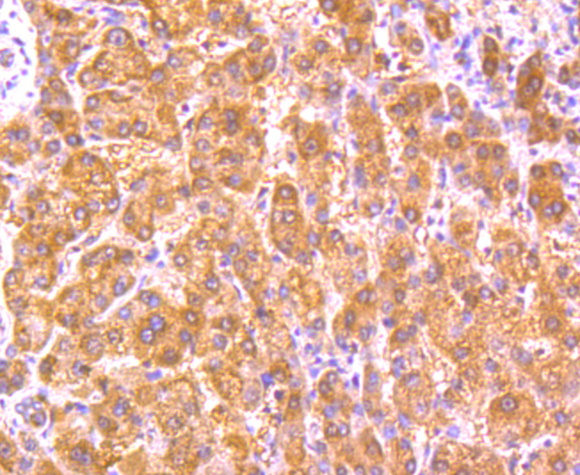
Fig5: Immunohistochemical analysis of paraffin-embedded human liver tissue using anti-GPX5 antibody. Counter stained with hematoxylin.Application
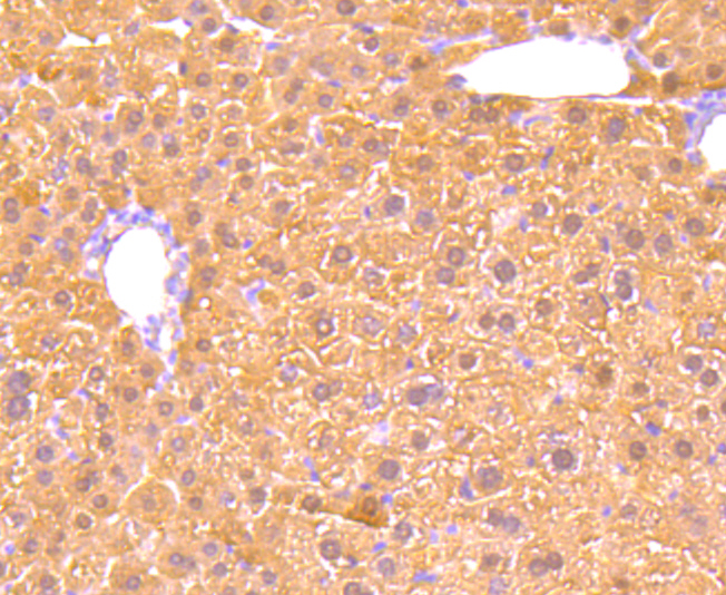
Fig6: Immunohistochemical analysis of paraffin-embedded mouse liver tissue using anti-GPX5 antibody. Counter stained with hematoxylin.Application
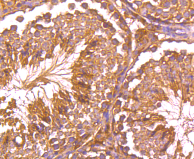
Fig7: Immunohistochemical analysis of paraffin-embedded mouse testis tissue using anti-GPX5 antibody. Counter stained with hematoxylin.| Product Name | GPX5 [5A1] |
|---|---|
| Antibody Type | Primary Antibodies |
| Antigen Alias | Arep arMEP24 EGLP Epididymal androgen related protein Epididymal secretory glutathione peroxidase epididymis secretory sperm binding protein Li 75p Epididymis specific glutathione peroxidase like protein Epididymis-specific glutathione peroxidase-like protein Glutathione peroxidase 5 Glutathione peroxidase 5 (epididymal androgen related protein) Glutathione peroxidase, epididymal GPx-5 GPX5 GPX5_HUMAN GSHPx-5 HEL-S-75p Major androgen regulated protein OTTHUMP00000017919 OTTHUMP00000214636 |
| Product description | Glutathione peroxidase (GPx) enzymes are generally selenium-containing tetrameric glycoproteins that help prevent lipid peroxidation of cell membranes. GPx enzymes reduce lipid hydroperoxides to alcohols, and reduce free hydrogen peroxide to water. GPx members are among the few proteins known in higher vertebrates to contain selenocysteine, which occurs at the active site of glutathione peroxidase and is coded by the nonsense (stop) codon TGA. There are eight GPx homologs (GPx-1-8). GPx-1, Gpx-2 and Gpx-3 exist as homotetramers. Gpx-4 has a high tendancy to form high molecular weight oligomers. GPx-1 plays an important role in the antioxidant defense of the vascular wall and neural cells in response to oxidative stress. GPx-2 is the major isoform in the lungs and its basal or inducible expression is dependent on Nrf2. GPx-3 is under regulation by hypoxic stress and the expression and deficiency of GPx-3 is associated with cardiovascular disease and stroke. GPx-5 is selenium-independent; it is bound to the acrosome of sperm, where it may protect sperm from premature acrosome reaction in the epididymis. |
| Immunogen | Peptide |
| Clonality | Monoclonal |
|---|---|
| Isotype | IgG1 |
| Host Species | Mouse |
| Tested Applications | WBIHCICC |
| WB:1:250~1:1000 IHC:1:20~1:100 ICC:1:100~1:200 Notes:Optimal dilutions/concentrations should be determined by the researcher. |
|
| Species Reactivity | HumanMouse |
| Concentration | 2mg/ml |
| Alternative Names | Arep
arMEP24
EGLP
Epididymal androgen related protein
Epididymal secretory glutathione peroxidase
epididymis secretory sperm binding protein Li 75p
Epididymis specific glutathione peroxidase like protein
Epididymis-specific glutathione peroxidase-like protein
Glutathione peroxidase 5
Glutathione peroxidase 5 (epididymal androgen related protein)
Glutathione peroxidase epididymal GPx-5 GPX5 GPX5_HUMAN GSHPx-5 HEL-S-75p Major androgen regulated protein OTTHUMP00000017919 OTTHUMP00000214636 |
|---|---|
| Molecular Weight(MW) | 22 kDa |
| Cellular Localization | Secreted. |
Application
Fig1: Western blot analysis of GPX5 on human sperm tissue lysate using anti-GPX5 antibody at 1/5,000 dilution.
Application
Fig2: Western blot analysis of GPX5 on mouse testis tissue lysate using anti-GPX5 antibody at 1/500 dilution.
Application
Fig3: ICC staining GPX5 (green) in 293T cells. The nuclear counter stain is DAPI (blue). Cells were fixed in paraformaldehyde, permeabilised with 0.25% Triton X100/PBS.
Application
Fig4: ICC staining GPX5 (green) in Hela cells. The nuclear counter stain is DAPI (blue). Cells were fixed in paraformaldehyde, permeabilised with 0.25% Triton X100/PBS.
Application
Fig5: Immunohistochemical analysis of paraffin-embedded human liver tissue using anti-GPX5 antibody. Counter stained with hematoxylin.
Application
Fig6: Immunohistochemical analysis of paraffin-embedded mouse liver tissue using anti-GPX5 antibody. Counter stained with hematoxylin.
Application
Fig7: Immunohistochemical analysis of paraffin-embedded mouse testis tissue using anti-GPX5 antibody. Counter stained with hematoxylin.| Positive Control | Mouse testis tissue lysate, human sperm tissue lysate, 293T, Hela, human liver tissue, mouse liver tissue, mouse testis tissue. |
|---|---|
| Application Notes | WB:1:250~1:1000 IHC:1:20~1:100 ICC:1:100~1:200 Notes:Optimal dilutions/concentrations should be determined by the researcher. |
| Form | Liquid |
|---|---|
| Storage Instructions | Store at +4℃ after thawing. Aliquot store at -20℃ or -80℃. Avoid repeated freeze / thaw cycles. |
| Storage Buffer | 1*TBS (pH7.4), 0.5%BSA, 50%Glycerol. Preservative: 0.05% Sodium Azide. |


