Application
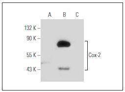
Fig1: Western blot analysis of Cox-2 expression in untreated (A), LPS treated (B) and LPS and Hydrocortisone treated (C) RAW 264.7 whole cell lysates. Note decreased LPS induction of Cox-2 expression in lane C.expression in lane C.Application
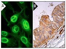
Fig2: Immunofluorescence staining of methanol-fixed NIH/3T3 cells showing membrane and nuclear envelope localization (A). Immunoperoxidase staining of formalin fixed, paraffin-embedded human gall bladder tissue showing cytoplasmic staining of glandular cells (B).| Product Name | COX2 |
|---|---|
| Antibody Type | Primary Antibodies |
| Antigen Alias | COX 2 antibody COX-2 antibody COX2 antibody Cyclooxygenase 2 antibody Cyclooxygenase 2b antibody Cyclooxygenase antibody Cyclooxygenase-2 antibody Cyclooxygenase2 antibody EC 1.14.99.1 antibody fj02a10 antibody Glucocorticoid-regulated inflammatory cyclooxygenase antibody Glucocorticoid-regulated inflammatory Prostaglandin G/H synthase antibody GRIPGHS antibody hCox 2 antibody Macrophage activation-associated marker protein P71/73 antibody OTTHUMP00000033524 antibody PES-2 antibody PGG/HS antibody PGH synthase 2 antibody PGH2_HUMAN antibody PGHS 2 antibody PGHS-2 antibody PGHS2 antibody PHS 2 antibody PHS II antibody PHS2 antibody Prostaglandin endoperoxide synthase 2 (prostaglandin G/H synthase and cyclooxygenase) antibody Prostaglandin endoperoxide synthase 2 antibody Prostaglandin G/H synthase 2 antibody Prostaglandin G/H synthase 2 precursor antibody Prostaglandin G/H synthase and cyclooxygenase antibody Prostaglandin G/H synthase antibody Prostaglandin H2 synthase 2 antibody prostaglandin-endoperoxide synthase 2 (prostaglandin G/H synthase and cyclooxygenase) antibody Prostaglandin-endoperoxide synthase 2 antibody PTGS2 antibody ptgs2a antibody TIS10 antibody TIS10 protein antibody unp1239 antibody wu:fj02a10 antibody |
| Product description | Prostaglandins are a diverse group of autocrine and paracrine hormones thatmediate many cellular and physiologic processes. Prostaglandin H2 (PGH2) isan intermediate molecule in formation of the prostaglandins. Cyclooxygenase-1(Cox-1) and cyclooxygenase-2 (Cox-2) are prostaglandin synthases that catalyzethe formation of PGH2 from arachidonic acid (AA). Cox-1 and Cox-2 areisozymes of prostaglandin-endoperoxidase synthase (PTGS). Cox-1 is constitutivelyexpressed in most tissues and is thought to serve in general “housekeeping”functions. Cox-2 is efficiently induced in migratory cells respondingto pro-inflammatory stimuli and is considered to be an important mediator ofinflammation. Both enzymes are targets for the nonsteroidal therapeutic antiinflammatorydrugs, NSAIDs. |
| Immunogen | peptide |
| Clonality | Monoclonal |
|---|---|
| Isotype | IgG |
| Host Species | Mouse |
| Tested Applications | WBIPIFIHCICC/IF |
| WB:1:100-1:1000 IP:1-2 μg per 100-500 μg of total protein (1 ml of cell lysate) IF:1:50-1:500 IHC:1:50-1:500 ELISA:1:30-1:3000 |
|
| Species Reactivity | HumanMouseRat |
| Concentration | 2mg/ml |
| Alternative Names | COX 2 antibody COX-2 antibody COX2 antibody Cyclooxygenase 2 antibody Cyclooxygenase 2b antibody Cyclooxygenase antibody Cyclooxygenase-2 antibody Cyclooxygenase2 antibody EC 1.14.99.1 antibody fj02a10 antibody Glucocorticoid-regulated inflammatory cyclooxygenase antibody Glucocorticoid-regulated inflammatory Prostaglandin G/H synthase antibody GRIPGHS antibody hCox 2 antibody Macrophage activation-associated marker protein P71/73 antibody OTTHUMP00000033524 antibody PES-2 antibody PGG/HS antibody PGH synthase 2 antibody PGH2_HUMAN antibody PGHS 2 antibody PGHS-2 antibody PGHS2 antibody PHS 2 antibody PHS II antibody PHS2 antibody Prostaglandin endoperoxide synthase 2 (prostaglandin G/H synthase and cyclooxygenase) antibody Prostaglandin endoperoxide synthase 2 antibody Prostaglandin G/H synthase 2 antibody Prostaglandin G/H synthase 2 precursor antibody Prostaglandin G/H synthase and cyclooxygenase antibody Prostaglandin G/H synthase antibody Prostaglandin H2 synthase 2 antibody prostaglandin-endoperoxide synthase 2 (prostaglandin G/H synthase and cyclooxygenase) antibody Prostaglandin-endoperoxide synthase 2 antibody PTGS2 antibody ptgs2a antibody TIS10 antibody TIS10 protein antibody unp1239 antibody wu:fj02a10 antibody |
|---|---|
| Molecular Weight(MW) | 70-72kDa |
| Cellular Localization | Microsome membrane, Endoplasmic reticulum membrane |
| SwissProt ID | P35354 |
|---|
Application
Fig1: Western blot analysis of Cox-2 expression in untreated (A), LPS treated (B) and LPS and Hydrocortisone treated (C) RAW 264.7 whole cell lysates. Note decreased LPS induction of Cox-2 expression in lane C.expression in lane C.
Application
Fig2: Immunofluorescence staining of methanol-fixed NIH/3T3 cells showing membrane and nuclear envelope localization (A). Immunoperoxidase staining of formalin fixed, paraffin-embedded human gall bladder tissue showing cytoplasmic staining of glandular cells (B).| Positive Control | RAW 264.7, NIH/3T3, human gall bladder tissue. |
|---|---|
| Application Notes | WB:1:100-1:1000 IP:1-2 μg per 100-500 μg of total protein (1 ml of cell lysate) IF:1:50-1:500 IHC:1:50-1:500 ELISA:1:30-1:3000 |
| Form | Liquid |
|---|---|
| Storage Instructions | Store at +4℃ |
| Storage Buffer | 1*TBS (pH7.4), 1%BSA, 40%Glycerol. Preservative: 0.05% Sodium Azide. |


