Application
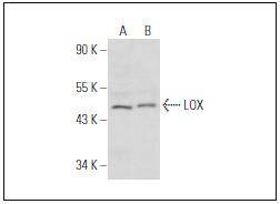
Fig1: Western blot analysis of LOX expression in WI-38 (A) and MCF7 (B) whole cell lysates.Application
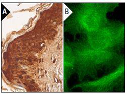
Fig2: Immunoperoxidase staining of formalin fixed, paraffin-embedded human skin tissue showing cytoplasmic and nuclear staining of epidermal cells (A). Immunofluorescence staining of formalinfixed HepG2 cells showing cell surface localization (B).| Product Name | LOX-1 [2G5] |
|---|---|
| Antibody Type | Primary Antibodies |
| Antigen Alias | lox antibody LYOX antibody LYOX_HUMAN antibody Lysyl oxidase antibody MGC105112 antibody Protein lysine 6 oxidase antibody Protein-lysine 6-oxidase antibody |
| Product description | The lysyl oxidase family of extracellular proteins includes LOX and four LOX-like enzymes, which are responsible for the deamination of peptidyl lysine residues of collagens and elastin. They also catalyze inter- and intra-crosslinking reactions. Overexpression of LOX may cause severe fibrotic degeneration due to its high resistance to degradative enzymes. Procollagen C-proteinase activity processes LOX from a precursor protein to a mature form. Activation of LOX occurs in normal developing and adult skin, and alterations in LOX expression and activity are associated with skin aging and senescence. LOX is crucial for development of the cardiovascular and respiratory systems. In addition, LOX plays a role in cancer, wound healing, as well as cell motility, chemotaxis and differentiation. |
| Immunogen | peptide |
| Clonality | Monoclonal |
|---|---|
| Isotype | IgG1 |
| Host Species | Mouse |
| Tested Applications | WBIPIFIHC-P |
| WB:1:100-1:1,000 IP:1-2 μg per 100-500 μg of total protein(1 ml of cell lysate) IF:1:50-1:500 IHC:1:50-1:500 |
|
| Species Reactivity | HumanMouseRat |
| Concentration | 2mg/ml |
| Alternative Names | lox antibody LYOX antibody LYOX_HUMAN antibody Lysyl oxidase antibody MGC105112 antibody Protein lysine 6 oxidase antibody Protein-lysine 6-oxidase antibody |
|---|---|
| Molecular Weight(MW) | 40 kDa |
| Cellular Localization | Secreted |
| SwissProt ID | P28300 |
|---|
Application
Fig1: Western blot analysis of LOX expression in WI-38 (A) and MCF7 (B) whole cell lysates.
Application
Fig2: Immunoperoxidase staining of formalin fixed, paraffin-embedded human skin tissue showing cytoplasmic and nuclear staining of epidermal cells (A). Immunofluorescence staining of formalinfixed HepG2 cells showing cell surface localization (B).| Positive Control | WI-38, MCF7, HepG2, HepG2. |
|---|---|
| Application Notes | WB:1:100-1:1,000 IP:1-2 μg per 100-500 μg of total protein(1 ml of cell lysate) IF:1:50-1:500 IHC:1:50-1:500 |
| Form | Liquid |
|---|---|
| Storage Instructions | Store at +4℃ |
| Storage Buffer | 1*TBS (pH7.4), 1%BSA, 40%Glycerol. Preservative: 0.05% Sodium Azide. |


