Application
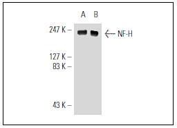
Fig1: Western blot analysis of NF-H expression in rat brain (A) and mouse brain (B) tissue extracts.Application
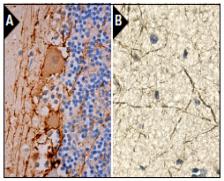
Fig2: Immunoperoxidase staining of formalin fixed, paraffin-embedded human cerebellum tissue showing membrane and cytoplasmic staining of Purkinje cells and neuropil staining in granular layer and molecular layer (A). Immunoperoxidase staining of formalin fixed, paraffin-embedded human cerebellum tissue showing membrane and cytoplasmic staining of Purkinje cells and neuropil staining in granular layer and molecular layer (B).| Product Name | NF-H [2G1] |
|---|---|
| Antibody Type | Primary Antibodies |
| Antigen Alias | 200 kDa neurofilament protein antibody CMT2CC antibody Nefh antibody Neurofilament heavy polypeptide 200kDa antibody Neurofilament heavy polypeptide antibody Neurofilament triplet H protein antibody NF H antibody NF-H antibody NFH antibody NFH_HUMAN antibody |
| Product description | Neurofilament-H (NF-H), for neurofilament heavy polypeptide, a member of the intermediate filament family, is a major component of neuronal cytoskeletons. Neurofilaments are dynamic structures; they contain phosphorylation sites for a large number of protein kinases, including protein kinase A, protein kinase C, cyclin-dependent kinase 5, extracellular signal regulated kinase, glycogen synthase kinase-3, and stress-activated protein kinase gamma. In addition to their role in the control of axon caliber, neurofilaments may affect other cytoskeletal elements, such as microtubules and Actin filaments. Changes in neurofilament phosphorylation or metabolism are frequently observed in neurodegenerative diseases, including amyotrophic lateral sclerosis (ALS), Parkinson's disease and Alzheimer's disease. |
| Immunogen | A neurofilament NF-H protein isolated from a cytoskeletal preparation from brain tissue homogenate of calf origin. |
| Clonality | Monoclonal |
|---|---|
| Isotype | IgG1 |
| Host Species | Mouse |
| Tested Applications | WBIPIFIHC-P |
| WB:1:500-1:1,000 IP:1-2 μg per 100-500 μg of total protein(1 ml of cell lysate) IF:1:50-1:500 IHC:1:50-1:500 |
|
| Species Reactivity | HumanMouseRat |
| Concentration | 2mg/ml |
| Alternative Names | 200 kDa neurofilament protein antibody CMT2CC antibody Nefh antibody Neurofilament heavy polypeptide 200kDa antibody Neurofilament heavy polypeptide antibody Neurofilament triplet H protein antibody NF H antibody NF-H antibody NFH antibody NFH_HUMAN antibody |
|---|---|
| Molecular Weight(MW) | 200 kDa |
| Cellular Localization | Cytoplasm, membrane |
| SwissProt ID | P12036 |
|---|
Application
Fig1: Western blot analysis of NF-H expression in rat brain (A) and mouse brain (B) tissue extracts.
Application
Fig2: Immunoperoxidase staining of formalin fixed, paraffin-embedded human cerebellum tissue showing membrane and cytoplasmic staining of Purkinje cells and neuropil staining in granular layer and molecular layer (A). Immunoperoxidase staining of formalin fixed, paraffin-embedded human cerebellum tissue showing membrane and cytoplasmic staining of Purkinje cells and neuropil staining in granular layer and molecular layer (B).| Positive Control | rat brain tissue, mouse brain tissue. |
|---|---|
| Application Notes | WB:1:500-1:1,000 IP:1-2 μg per 100-500 μg of total protein(1 ml of cell lysate) IF:1:50-1:500 IHC:1:50-1:500 |
| Form | Liquid |
|---|---|
| Storage Instructions | Store at +4℃ |
| Storage Buffer | 1*TBS (pH7.4), 1%BSA, 40%Glycerol. Preservative: 0.05% Sodium Azide. |


