Application
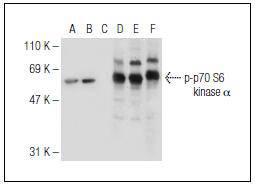
Fig1: Western blot analysis of p70 S6 kinase α phosphorylation in untreated (A,D), insulin treated (B,E) and insulin treated and lambda protein phosphatase treated (C,F) HEK293 whole cell lysates. Antibodies tested include p-p70 S6 kinase α (A-6) (A,B,C) and p70 S6 kinase α (C-18) (D,E,F).Application
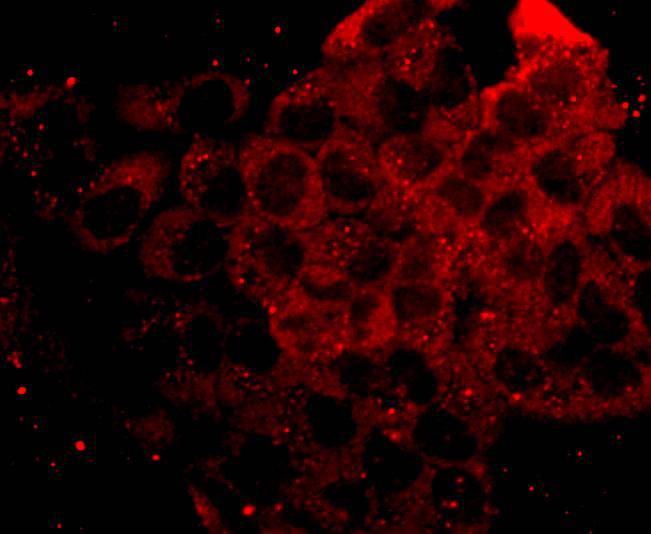
Fig2: Immunofluorescence staining of anisomycin-treated, methanol-fixed NIH/3T3 cells, showing cytoplasmic and nuclear localization of activated p70 S6 kinase α (A). Immunoperoxidase staining of formalin fixed, paraffin-embedded human esophagus tissue showing nuclear staining of squamous epithelial cells (B).| Product Name | Phospho-p70 S6 Kinase (Ser 411) [2G1] |
|---|---|
| Antibody Type | Primary Antibodies |
| Antigen Alias | 70 kDa ribosomal protein S6 kinase 2 antibody EC 2.7.11.1 antibody KS6B2_HUMAN antibody p70 beta antibody p70 ribosomal S6 kinase beta antibody p70 S6 kinase beta antibody p70 S6K-beta antibody p70 S6KB antibody p70 S6Kbeta antibody p70(S6K) beta antibody p70-beta antibody p70-S6K 2 antibody P70S6K2 antibody p70S6Kb antibody Ribosomal protein S6 kinase 70kDa, polypeptide 2 antibody Ribosomal protein S6 kinase beta 2 antibody Ribosomal protein S6 kinase beta-2 antibody Rps6kb2 antibody S6 kinase related kinase antibody S6 kinase-related kinase antibody S6K beta 2 antibody S6K beta antibody S6K-beta antibody S6K-beta-2 antibody S6K2 antibody Serine/threonine protein kinase 14 beta antibody Serine/threonine-protein kinase 14B antibody SRK antibody STK14B antibody |
| Product description | In studies to elucidate key regulatory pathways in signal transduction, several protein serine/threonine (Ser/Thr) kinases have been identified. Included among such kinases are two distinct families of 40S ribosomal protein S6 Ser/Thr kinases present in somatic animal cells, designated p70 S6 kinase and p90 Rsk kinase. p90 Rsk kinase is maximally activated within minutes of addition of growth factors or phorbol ester to cultured cells followed by activation of p70 S6 kinase. Both enzymes are regulated by serine/threonine phosphorylation, suggesting that specific kinases may exist upstream in the signaling pathway that regulate these kinases. In fact, evidence suggests that one such family of activating enzymes includes the members of the ERK MAP kinase family. The ERK MAP kinases are, in turn, regulated by phosphorylation at threonine and tyrosine residues by a protein kinase designated MEK. |
| Immunogen | A sequence containing Ser 411 phosphorylated p70 S6 kinase α isoform 2 of human origin. |
| Modification | p-Ser 411 |
| Clonality | Monoclonal |
|---|---|
| Isotype | IgG1 |
| Host Species | Mouse |
| Tested Applications | WBIPIFIHC-P |
| WB:1:100-1:1,000 IP:1-2 μg per 100-500 μg of total protein(1 ml of cell lysate) IF:1:50-1:500 IHC:1:50-1:500 |
|
| Species Reactivity | HumanMouseRat |
| Concentration | 2mg/ml |
| Alternative Names | 70 kDa ribosomal protein S6 kinase 2 antibody
EC 2.7.11.1 antibody
KS6B2_HUMAN antibody
p70 beta antibody
p70 ribosomal S6 kinase beta antibody
p70 S6 kinase beta antibody
p70 S6K-beta antibody
p70 S6KB antibody
p70 S6Kbeta antibody
p70(S6K) beta antibody
p70-beta antibody
p70-S6K 2 antibody
P70S6K2 antibody
p70S6Kb antibody
Ribosomal protein S6 kinase 70kDa polypeptide 2 antibody Ribosomal protein S6 kinase beta 2 antibody Ribosomal protein S6 kinase beta-2 antibody Rps6kb2 antibody S6 kinase related kinase antibody S6 kinase-related kinase antibody S6K beta 2 antibody S6K beta antibody S6K-beta antibody S6K-beta-2 antibody S6K2 antibody Serine/threonine protein kinase 14 beta antibody Serine/threonine-protein kinase 14B antibody SRK antibody STK14B antibody |
|---|---|
| Molecular Weight(MW) | 70kDa |
| Cellular Localization | Cytoplasm, Nucleus |
| SwissProt ID | Q9UBS0 |
|---|
Application
Fig1: Western blot analysis of p70 S6 kinase α phosphorylation in untreated (A,D), insulin treated (B,E) and insulin treated and lambda protein phosphatase treated (C,F) HEK293 whole cell lysates. Antibodies tested include p-p70 S6 kinase α (A-6) (A,B,C) and p70 S6 kinase α (C-18) (D,E,F).
Application
Fig2: Immunofluorescence staining of anisomycin-treated, methanol-fixed NIH/3T3 cells, showing cytoplasmic and nuclear localization of activated p70 S6 kinase α (A). Immunoperoxidase staining of formalin fixed, paraffin-embedded human esophagus tissue showing nuclear staining of squamous epithelial cells (B).| Positive Control | HEK293, NIH/3T3, human esophagus tissue. |
|---|---|
| Application Notes | WB:1:100-1:1,000 IP:1-2 μg per 100-500 μg of total protein(1 ml of cell lysate) IF:1:50-1:500 IHC:1:50-1:500 |
| Form | Liquid |
|---|---|
| Storage Instructions | Store at +4℃ |
| Storage Buffer | 1*TBS (pH7.4), 1%BSA, 40%Glycerol. Preservative: 0.05% Sodium Azide. |


