




WB
Western blot analysis using Nrf2 antibody against Hepg2 (1) cell lysate.IHC
Immunohistochemical analysis of paraffin-embedded Human breast cancer tissues using Nrf2 antibody with DAB staining.IHC
Immunohistochemical analysis of paraffin-embedded Human esophageal tissues using Nrf2 antibody with DAB staining.IHC
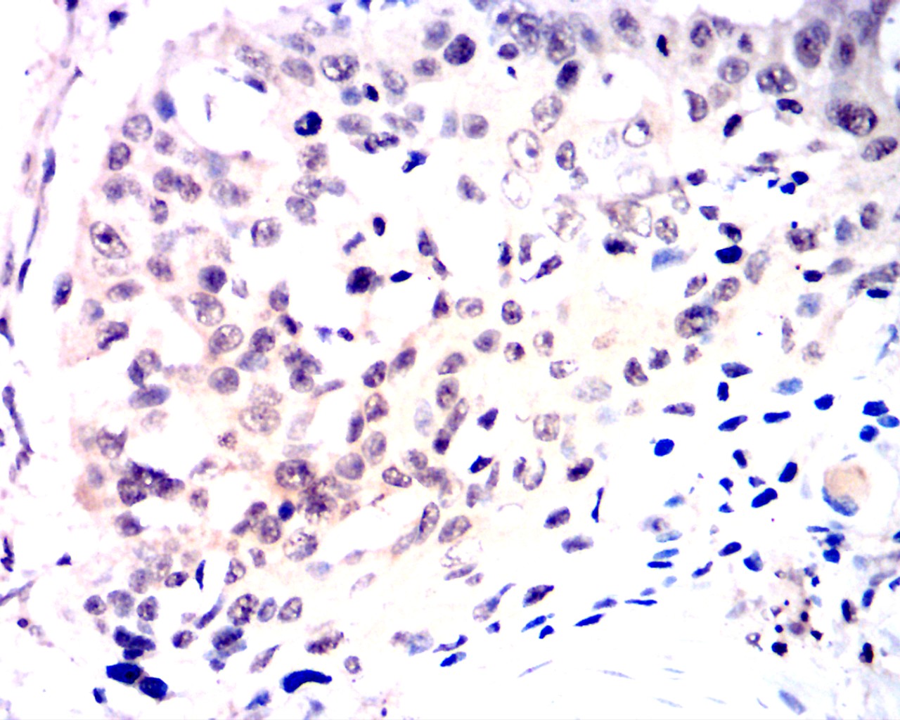
Immunohistochemical analysis of paraffin-embedded Human esophageal cancer tissues using Nrf2 antibody with DAB staining.IHC
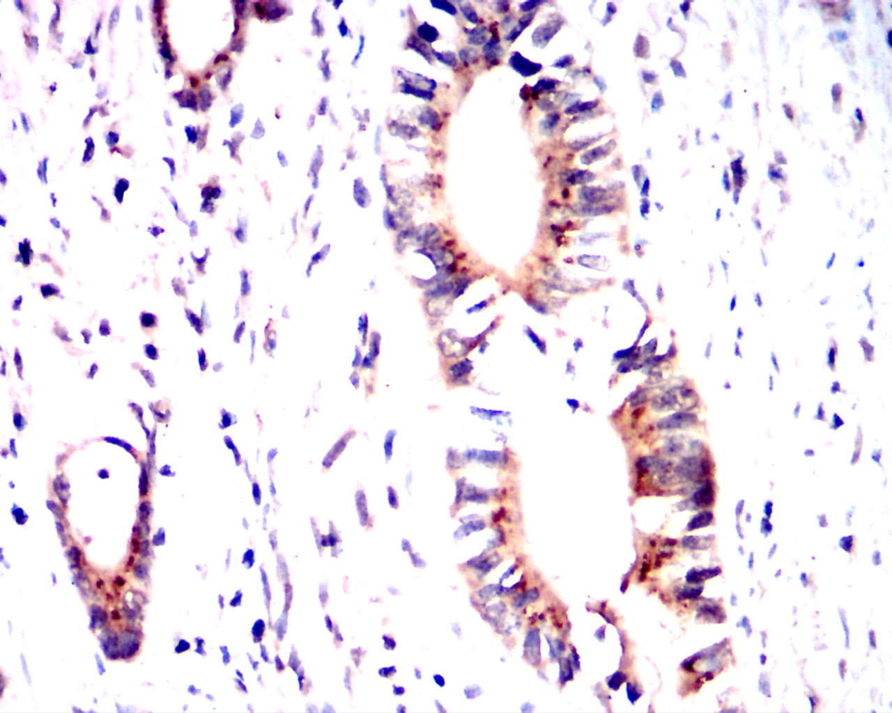
Immunohistochemical analysis of paraffin-embedded Human rectal cancer tissues using Nrf2 antibody with DAB staining.WB
(B–D) Application of Western blot (B, C) and qT-PCR (D) to ascertain the protein and mRNA expression levels, respectively, in maternal placenta, liver, and pancreas tissues.WB
(B, C) Western blotting analysis was used to detect theNrf2 and CBR1 expression level at week 0 and 16 in offspring rat liver (B) and pancreas (C).WB
(A) BRL cells were exposed to 25-hydroxyvitaminD (VD3) at 0, 10, 100, 1,000 nM for 24 h, and thenNrf2 levels were evaluated by Western blot.WB
(b–d) Western blot (b,c) and qRT-PCR (d) to ascertain the protein and mRNA expression levels in maternal placenta tissues.| Product Name | Anti-Nrf2 antibody-poly-HRP |
|---|---|
| Antibody Type | Primary Antibodies |
| Product description |
|
| Immunogen | Polypeptide |
| Conjugation | poly-HRP |
| Clonality | Polyclonal |
|---|---|
| Isotype | IgG |
| Host Species | Rabbit |
| Tested Applications | WBIHC |
| WB:1:200-1:2000 IHC:1:100-1:1000 |
|
| Species Reactivity | HumanMouseRat |
| Concentration | 1mg/ml |
| Purification | Protein A |
| Gene Symbol | NFE2L2 |
|---|---|
| Gene Synonyms | IMDDHH Nrf-2 NRF2 |
| Gene Full Name | NFE2 like bZIP transcription factor 2 |
| Gene Summary | This gene encodes a transcription factor which is a member of a small family of basic leucine zipper (bZIP) proteins. The encoded transcription factor regulates genes which contain antioxidant response elements (ARE) in their promoters; many of these genes encode proteins involved in response to injury and inflammation which includes the production of free radicals. Multiple transcript variants encoding different isoforms have been characterized for this gene. [provided by RefSeq, Sep 2015] |
| Alternative Names | E4TF1-60 E4TF1A NF-E2-like basic leucine zipper transcriptional activator NF-E2-related factor 2 NFE2-related factor 2 NFT2 NRF2 NRF2A nuclear factor erythroid-derived 2-like 2 nuclear factor erythroid 2 like 2 nuclear factor erythroid 2-like 2 nuclear factor erythroid derived 2 like 2 transcription factor |
| Molecular Weight(MW) | 68 kDa |
| Cellular Localization | Cytosol;Nucleus |
| SwissProt ID | Q16236 |
|---|
WB
Western blot analysis using Nrf2 antibody against Hepg2 (1) cell lysate.
IHC
Immunohistochemical analysis of paraffin-embedded Human breast cancer tissues using Nrf2 antibody with DAB staining.
IHC
Immunohistochemical analysis of paraffin-embedded Human esophageal tissues using Nrf2 antibody with DAB staining.
IHC
Immunohistochemical analysis of paraffin-embedded Human esophageal cancer tissues using Nrf2 antibody with DAB staining.
IHC
Immunohistochemical analysis of paraffin-embedded Human rectal cancer tissues using Nrf2 antibody with DAB staining.
WB
(B–D) Application of Western blot (B, C) and qT-PCR (D) to ascertain the protein and mRNA expression levels, respectively, in maternal placenta, liver, and pancreas tissues.
WB
(B, C) Western blotting analysis was used to detect theNrf2 and CBR1 expression level at week 0 and 16 in offspring rat liver (B) and pancreas (C).
WB
(A) BRL cells were exposed to 25-hydroxyvitaminD (VD3) at 0, 10, 100, 1,000 nM for 24 h, and thenNrf2 levels were evaluated by Western blot.
WB
(b–d) Western blot (b,c) and qRT-PCR (d) to ascertain the protein and mRNA expression levels in maternal placenta tissues.| Application Notes | WB:1:200-1:2000 IHC:1:100-1:1000 |
|---|
| Form | Liquid |
|---|---|
| Storage Instructions | Shipped at 4°C. Store at +4°C short term (1-2 weeks). Store at -20°C long term. Avoid freeze / thaw cycle. |
| Storage Buffer | Purified antibody in PBS with 0.05% PC300. |


