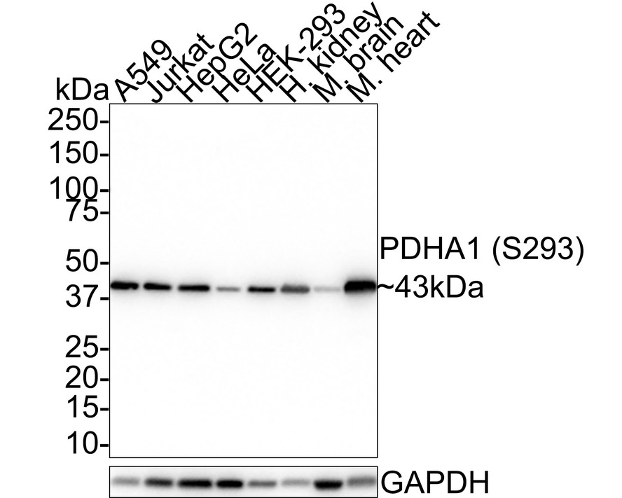
WB
Western blot analysis of Phospho-PDHA1 (S293) on different lysates with Rabbit anti-Phospho-PDHA1 (S293) antibody at 1/1,000 dilution. Lane 1: A549 cell lysate (20 µg/Lane) Lane 2: Jurkat cell lysate (20 µg/Lane) Lane 3: HepG2 cell lysate (20 µg/Lane) Lane 4: HeLa cell lysate (20 µg/Lane) Lane 5: HEK-293 cell lysate (20 µg/Lane) Lane 6: Human kidney tissue lysate (40 µg/Lane) Lane 7: Mouse brain tissue lysate (40 µg/Lane) Lane 8: Mouse heart tissue lysate (40 µg/Lane) Predicted band size: 43 kDa Observed band size: 43 kDa Exposure time: 39 seconds; 4-20% SDS-PAGE gel. Proteins were transferred to a PVDF membrane and blocked with 5% NFDM/TBST for 1 hour at room temperature. The primary antibody at 1/1,000 dilution was used in 5% NFDM/TBST at room temperature for 2 hours. Goat Anti-Rabbit IgG - HRP Secondary Antibody at 1:100,000 dilution was used for 1 hour at room temperature.IHC
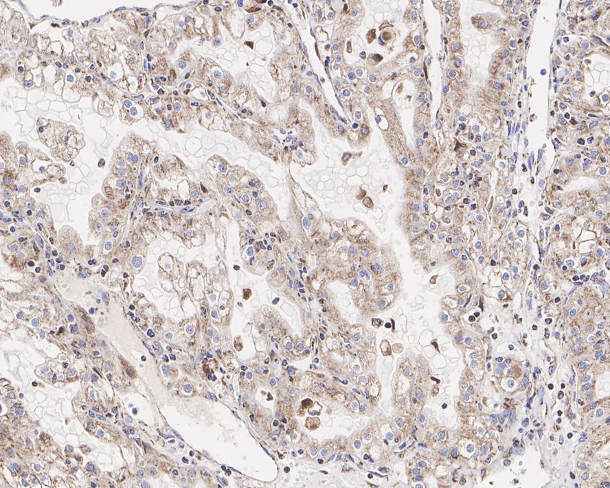
Immunohistochemical analysis of paraffin-embedded human renal clear cell carcinoma tissue with Rabbit anti-Phospho-PDHA1 (S293) antibody at 1/500 dilution. The section was pre-treated using heat mediated antigen retrieval with Tris-EDTA buffer (pH 9.0) for 20 minutes. The tissues were blocked in 1% BSA for 20 minutes at room temperature, washed with ddH2O and PBS, and then probed with the primary antibody at 1/500 dilution for 1 hour at room temperature. The detection was performed using an HRP conjugated compact polymer system. DAB was used as the chromogen. Tissues were counterstained with hematoxylin and mounted with DPX.ICC/IF
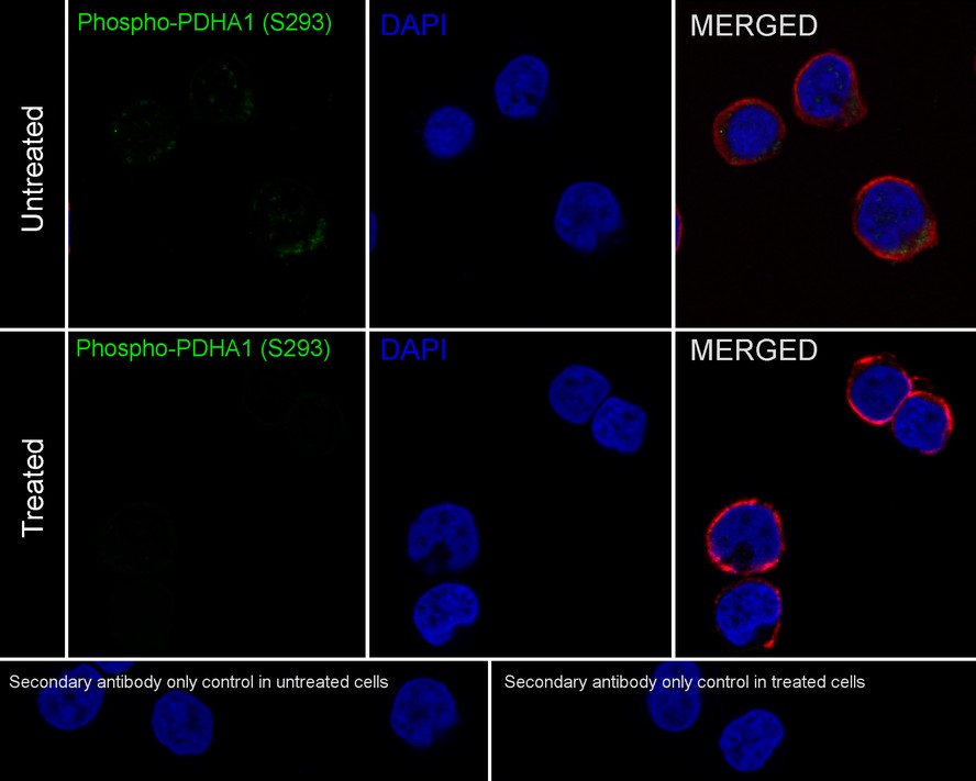
Immunocytochemistry analysis of Jurkat cells treated with or without λpp labeling Phospho-PDHA1 (S293) with Rabbit anti-Phospho-PDHA1 (S293) antibody at 1/100 dilution. Cells were fixed in 4% paraformaldehyde for 10 minutes at 37 ℃, permeabilized with 0.05% Triton X-100 in PBS for 20 minutes, and then blocked with 2% negative goat serum for 30 minutes at room temperature. Cells were then incubated with Rabbit anti-Phospho-PDHA1 (S293) antibody at 1/100 dilution in 2% negative goat serum overnight at 4 ℃. Goat Anti-Rabbit IgG H&L (iFluor™ 488) was used as the secondary antibody at 1/1,000 dilution. Nuclear DNA was labelled in blue with DAPI. Beta tubulin (red) was stained at 1/200 dilution overnight at +4℃. Goat Anti-Mouse IgG H&L (iFluor™ 594) was used as the secondary antibody at 1/1,000 dilution.| Product Name | Phospho-PDHA1 (S293) Recombinant Rabbit Monoclonal Antibody |
|---|---|
| Antibody Type | Primary Antibodies |
| Immunogen | Synthetic phosphopeptide corresponding to residues surrounding Ser293 of human PDHA1 protein. |
| Clonality | Monoclonal |
|---|---|
| Isotype | IgG |
| Host Species | Rabbit |
| Tested Applications | ICC/IFIHCWB |
| WB:1:1000 IHC:1:500-1:2000 ICC:1:100 |
|
| Species Reactivity | HumanMouseRat |
| Concentration | 1mg/ml |
| Purification | Protein A |
| Gene Symbol | PDHA1 |
|---|---|
| Gene Synonyms | PDHA PDHAD PHE1A E1alpha PDHCE1A |
| Gene Full Name | pyruvate dehydrogenase E1 subunit alpha 1 |
| Gene Summary | The pyruvate dehydrogenase (PDH) complex is a nuclear-encoded mitochondrial multienzyme complex that catalyzes the overall conversion of pyruvate to acetyl-CoA and CO(2), and provides the primary link between glycolysis and the tricarboxylic acid (TCA) cycle. The PDH complex is composed of multiple copies of three enzymatic components: pyruvate dehydrogenase (E1), dihydrolipoamide acetyltransferase (E2) and lipoamide dehydrogenase (E3). The E1 enzyme is a heterotetramer of two alpha and two beta subunits. This gene encodes the E1 alpha 1 subunit containing the E1 active site, and plays a key role in the function of the PDH complex. Mutations in this gene are associated with pyruvate dehydrogenase E1-alpha deficiency and X-linked Leigh syndrome. Alternatively spliced transcript variants encoding different isoforms have been found for this gene.[provided by RefSeq, Mar 2010] |
| Molecular Weight(MW) | 43kDa |
| Cellular Localization | Mitochondrion matrix. |
WB
Western blot analysis of Phospho-PDHA1 (S293) on different lysates with Rabbit anti-Phospho-PDHA1 (S293) antibody at 1/1,000 dilution. Lane 1: A549 cell lysate (20 µg/Lane) Lane 2: Jurkat cell lysate (20 µg/Lane) Lane 3: HepG2 cell lysate (20 µg/Lane) Lane 4: HeLa cell lysate (20 µg/Lane) Lane 5: HEK-293 cell lysate (20 µg/Lane) Lane 6: Human kidney tissue lysate (40 µg/Lane) Lane 7: Mouse brain tissue lysate (40 µg/Lane) Lane 8: Mouse heart tissue lysate (40 µg/Lane) Predicted band size: 43 kDa Observed band size: 43 kDa Exposure time: 39 seconds; 4-20% SDS-PAGE gel. Proteins were transferred to a PVDF membrane and blocked with 5% NFDM/TBST for 1 hour at room temperature. The primary antibody at 1/1,000 dilution was used in 5% NFDM/TBST at room temperature for 2 hours. Goat Anti-Rabbit IgG - HRP Secondary Antibody at 1:100,000 dilution was used for 1 hour at room temperature.
IHC
Immunohistochemical analysis of paraffin-embedded human renal clear cell carcinoma tissue with Rabbit anti-Phospho-PDHA1 (S293) antibody at 1/500 dilution. The section was pre-treated using heat mediated antigen retrieval with Tris-EDTA buffer (pH 9.0) for 20 minutes. The tissues were blocked in 1% BSA for 20 minutes at room temperature, washed with ddH2O and PBS, and then probed with the primary antibody at 1/500 dilution for 1 hour at room temperature. The detection was performed using an HRP conjugated compact polymer system. DAB was used as the chromogen. Tissues were counterstained with hematoxylin and mounted with DPX.
ICC/IF
Immunocytochemistry analysis of Jurkat cells treated with or without λpp labeling Phospho-PDHA1 (S293) with Rabbit anti-Phospho-PDHA1 (S293) antibody at 1/100 dilution. Cells were fixed in 4% paraformaldehyde for 10 minutes at 37 ℃, permeabilized with 0.05% Triton X-100 in PBS for 20 minutes, and then blocked with 2% negative goat serum for 30 minutes at room temperature. Cells were then incubated with Rabbit anti-Phospho-PDHA1 (S293) antibody at 1/100 dilution in 2% negative goat serum overnight at 4 ℃. Goat Anti-Rabbit IgG H&L (iFluor™ 488) was used as the secondary antibody at 1/1,000 dilution. Nuclear DNA was labelled in blue with DAPI. Beta tubulin (red) was stained at 1/200 dilution overnight at +4℃. Goat Anti-Mouse IgG H&L (iFluor™ 594) was used as the secondary antibody at 1/1,000 dilution.| Application Notes | WB:1:1000 IHC:1:500-1:2000 ICC:1:100 |
|---|
| Form | Liquid |
|---|---|
| Storage Instructions | Store at +4℃ after thawing. Aliquot store at -20℃. Avoid repeated freeze / thaw cycles. |
| Storage Buffer | 1*TBS (pH7.4), 0.05% BSA, 40% Glycerol. Preservative: 0.05% Sodium Azide. |


 Data sheet for OM643078