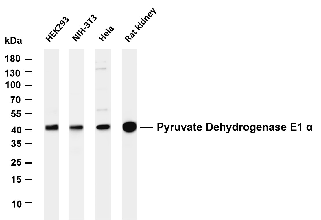
WB
Various whole cell lysates were separated by 4-20% SDS-PAGE, and the membrane was blotted with anti-Pyruvate Dehydrogenase E1 α antibody. The HRP-conjugated Goat anti-Rabbit IgG(H + L) antibody was used to detect the antibody. Lane 1: HEK293, Lane 2: NIH-3T3, Lane 3: Hela, Lane 4: Rat kidney.IHC
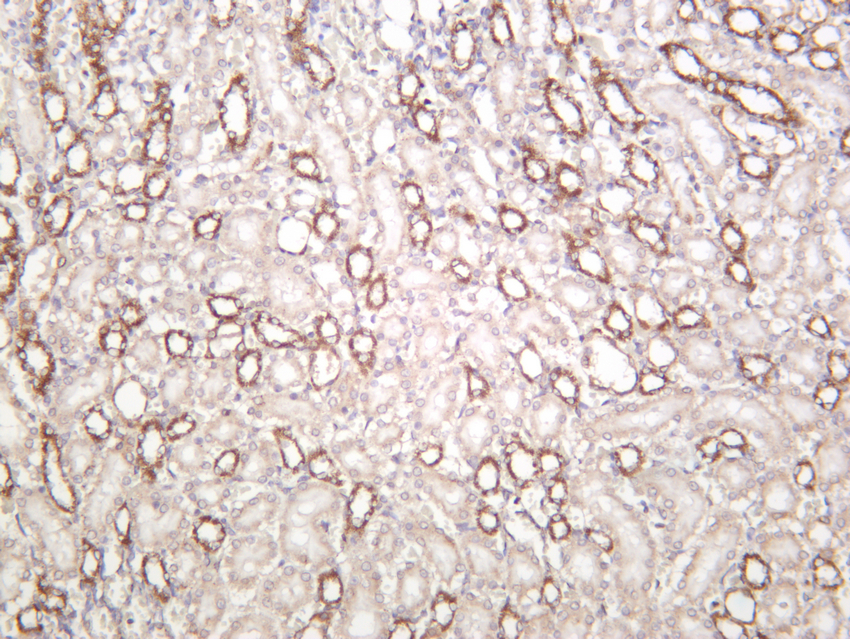
Rat kidney was stained with anti-Pyruvate Dehydrogenase E1 α rabbit antibody.IHC
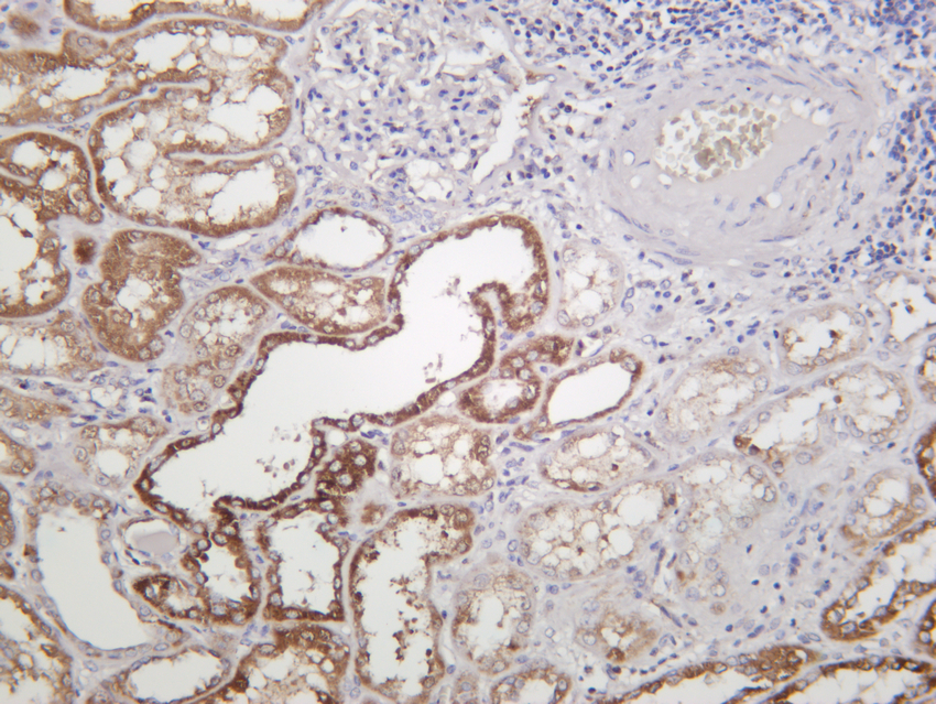
Human kidney was stained with anti-Pyruvate Dehydrogenase E1 α rabbit antibody.ICC/IF
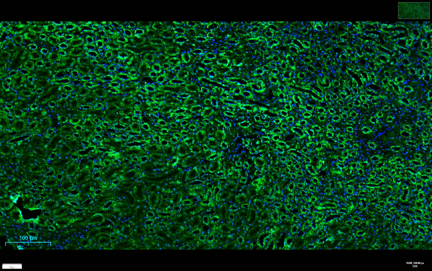
Mouse kidney was stained with anti-Pyruvate Dehydrogenase E1 α rabbit antibody.ICC/IF
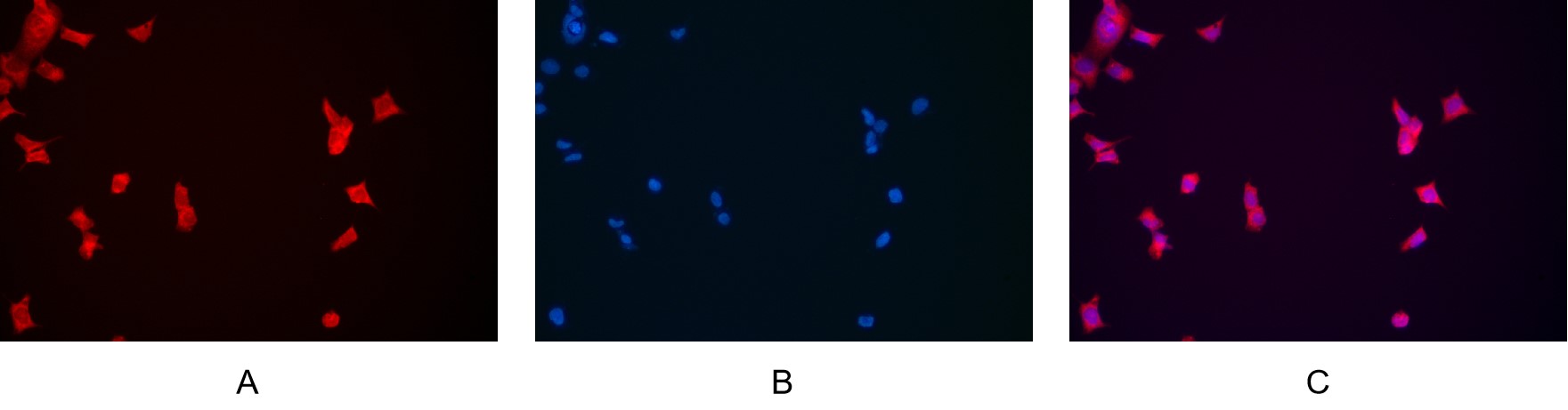
Immunofluorescence analysis of HEK293. Picture A: Pyruvate Dehydrogenase E1 alpha antibody (red). Picture B: DAPI (blue). Picture C: Merge of A+B.| Product Name | Pyruvate Dehydrogenase E1α Rabbit mAb |
|---|---|
| Antibody Type | Primary Antibodies |
| Immunogen | Pyruvate dehydrogenase E1 component subunit alpha somatic form mitochondrial. |
| Clonality | monoclonal |
|---|---|
| Isotype | IgG |
| Host Species | Rabbit |
| Tested Applications | ICC/IFIF-PIHCWB |
| WB:1:1000-1:5000 IHC:1:200-1:1000 ICC/IF:1:200-1:1000 IF-P:1:200-1:1000 |
|
| Species Reactivity | HumanMouseRat |
| Concentration | 1mg/ml |
| Purification | Protein A |
| Gene Symbol | PDHA1 |
|---|---|
| Gene Synonyms | PDHA PDHAD PHE1A E1alpha PDHCE1A |
| Gene Full Name | pyruvate dehydrogenase E1 subunit alpha 1 |
| Gene Summary | The pyruvate dehydrogenase (PDH) complex is a nuclear-encoded mitochondrial multienzyme complex that catalyzes the overall conversion of pyruvate to acetyl-CoA and CO(2), and provides the primary link between glycolysis and the tricarboxylic acid (TCA) cycle. The PDH complex is composed of multiple copies of three enzymatic components: pyruvate dehydrogenase (E1), dihydrolipoamide acetyltransferase (E2) and lipoamide dehydrogenase (E3). The E1 enzyme is a heterotetramer of two alpha and two beta subunits. This gene encodes the E1 alpha 1 subunit containing the E1 active site, and plays a key role in the function of the PDH complex. Mutations in this gene are associated with pyruvate dehydrogenase E1-alpha deficiency and X-linked Leigh syndrome. Alternatively spliced transcript variants encoding different isoforms have been found for this gene.[provided by RefSeq, Mar 2010] |
| Molecular Weight(MW) | 43kDa |
| Cellular Localization | Mitochondrion matrix. |
WB
Various whole cell lysates were separated by 4-20% SDS-PAGE, and the membrane was blotted with anti-Pyruvate Dehydrogenase E1 α antibody. The HRP-conjugated Goat anti-Rabbit IgG(H + L) antibody was used to detect the antibody. Lane 1: HEK293, Lane 2: NIH-3T3, Lane 3: Hela, Lane 4: Rat kidney.
IHC
Rat kidney was stained with anti-Pyruvate Dehydrogenase E1 α rabbit antibody.
IHC
Human kidney was stained with anti-Pyruvate Dehydrogenase E1 α rabbit antibody.
ICC/IF
Mouse kidney was stained with anti-Pyruvate Dehydrogenase E1 α rabbit antibody.
ICC/IF
Immunofluorescence analysis of HEK293. Picture A: Pyruvate Dehydrogenase E1 alpha antibody (red). Picture B: DAPI (blue). Picture C: Merge of A+B.| Application Notes | WB:1:1000-1:5000 IHC:1:200-1:1000 ICC/IF:1:200-1:1000 IF-P:1:200-1:1000 |
|---|
| Form | Liquid |
|---|---|
| Storage Instructions | -15°C to -25°C/1 year(Do not lower than -25°C) |
| Storage Buffer | PBS, 50% glycerol, 0.05% Proclin 300, 0.05%BSA |


 Data sheet for OM644237