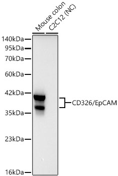
WB
Western blot analysis of various lysates using CD326/EpCAM Rabbit mAb at 1:3000 dilution. Secondary antibody: HRP-conjugated Goat anti-Rabbit IgG (H+L) at 1:10000 dilution. Lysates/proteins: 25ug per lane. Blocking buffer: 3% nonfat dry milk in TBST. Detection: ECL Basic Kit . Negative control (NC): C2C12.IHC
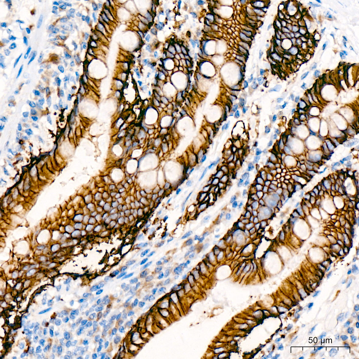
Immunohistochemistry analysis of paraffin-embedded Human small intestine tissue using CD326/EpCAM Rabbit mAb at a dilution of 1:1000 (40x lens). High pressure antigen retrieval was performed with 0.01 M citrate buffer (pH 6.0) prior to IHC staining. Exposure time: 20s.IF-P
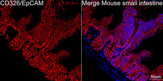
Confocal imaging of paraffin-embedded Mouse small intestine tissue using CD326/EpCAM Rabbit mAb (dilution 1:200) followed by a further incubation with Cy3 Goat Anti-Rabbit IgG (H+L) (dilution 1:500) (Red). DAPI was used for nuclear staining (Blue). Perform high pressure antigen retrieval with 0.01 M citrate buffer (pH 6.0) prior to IF staining. objective: 40x.FC
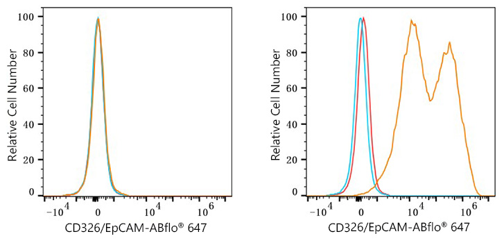
Flow cytometry: 1X10^6 293T cells (negative control,left) and 293T (Transfection,right) cells were surface-stained with CD326/EpCAM Rabbit mAb (2ug/mL,orange line) or ABflo® 647 Rabbit IgG isotype control (5ul/Test,blue line), followed by Alexa Fluor® 647 conjugated goat anti-rabbit pAb staining. Non-fluorescently stained cells were used as blank control (red line).| Product Name | CD326/EpCAM Rabbit mAb |
|---|---|
| Antibody Type | Primary Antibodies |
| Immunogen | Recombinant fusion protein containing a sequence corresponding to amino acids 24-266 of mouse CD326/EpCAM (NP_032558.2). |
| Clonality | Monoclonal |
|---|---|
| Isotype | IgG |
| Host Species | Rabbit |
| Tested Applications | FCIF-PIHCWB |
| WB:1:3000-1:12000 IHC:1:500-1:5000 IF-P:1:200-1:2000 FC:1:500-1:1000 |
|
| Species Reactivity | HumanMouseRat |
| Concentration | 1mg/ml |
| Purification | Affinity purified |
| Gene Symbol | Epcam |
|---|---|
| Gene Synonyms | EGP Ly74 gp40 CD326 EGP-2 TROP1 Egp314 Ep-CAM EpCAM1 Tacsd1 GA733-2 Tacstd1 |
| Gene Full Name | epithelial cell adhesion molecule |
| Gene Summary | Predicted to enable cadherin binding activity involved in cell-cell adhesion. Involved in ureteric bud development. Located in cell surface. Is expressed in several structures, including alimentary system; central nervous system; genitourinary system; respiratory system; and sensory organ. Used to study congenital diarrhea 5 with tufting enteropathy. Human ortholog(s) of this gene implicated in congenital diarrhea 5 with tufting enteropathy and hereditary nonpolyposis colorectal cancer type 8. Orthologous to human EPCAM (epithelial cell adhesion molecule). [provided by Alliance of Genome Resources, Nov 2024] |
| Molecular Weight(MW) | 35kDa |
| Cellular Localization | Lateral cell membrane. |
WB
Western blot analysis of various lysates using CD326/EpCAM Rabbit mAb at 1:3000 dilution. Secondary antibody: HRP-conjugated Goat anti-Rabbit IgG (H+L) at 1:10000 dilution. Lysates/proteins: 25ug per lane. Blocking buffer: 3% nonfat dry milk in TBST. Detection: ECL Basic Kit . Negative control (NC): C2C12.
IHC
Immunohistochemistry analysis of paraffin-embedded Human small intestine tissue using CD326/EpCAM Rabbit mAb at a dilution of 1:1000 (40x lens). High pressure antigen retrieval was performed with 0.01 M citrate buffer (pH 6.0) prior to IHC staining. Exposure time: 20s.
IF-P
Confocal imaging of paraffin-embedded Mouse small intestine tissue using CD326/EpCAM Rabbit mAb (dilution 1:200) followed by a further incubation with Cy3 Goat Anti-Rabbit IgG (H+L) (dilution 1:500) (Red). DAPI was used for nuclear staining (Blue). Perform high pressure antigen retrieval with 0.01 M citrate buffer (pH 6.0) prior to IF staining. objective: 40x.
FC
Flow cytometry: 1X10^6 293T cells (negative control,left) and 293T (Transfection,right) cells were surface-stained with CD326/EpCAM Rabbit mAb (2ug/mL,orange line) or ABflo® 647 Rabbit IgG isotype control (5ul/Test,blue line), followed by Alexa Fluor® 647 conjugated goat anti-rabbit pAb staining. Non-fluorescently stained cells were used as blank control (red line).| Application Notes | WB:1:3000-1:12000 IHC:1:500-1:5000 IF-P:1:200-1:2000 FC:1:500-1:1000 |
|---|
| Form | Liquid |
|---|---|
| Storage Instructions | Store at -20℃. Avoid freeze / thaw cycles. |
| Storage Buffer | Buffer: PBS with 0.09% Sodium azide,0.05% BSA,50% glycerol,pH7.3. |


 Data sheet for OM643202