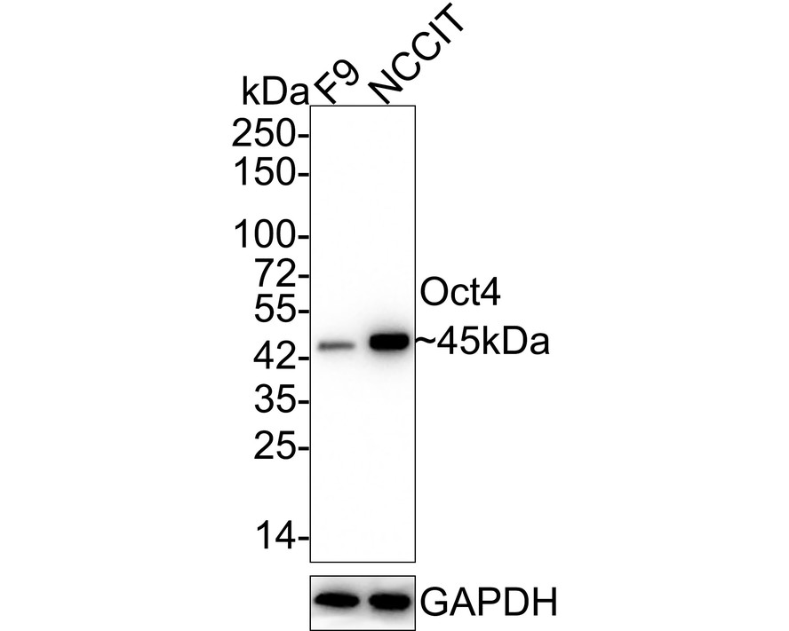
WB
Western blot analysis of Oct4 on different lysates with Rabbit anti-Oct4 antibody at 1/1,000 dilution. Lane 1: F9 cell lysate, Lane 2: NCCIT cell lysate, Lysates/proteins at 20 µg/Lane. Exposure time: 1 minute 59 seconds; 4-20% SDS-PAGE gel. Proteins were transferred to a PVDF membrane and blocked with 5% NFDM/TBST for 1 hour at room temperature. The primary antibody at 1/1,000 dilution was used in 5% NFDM/TBST at 4℃ overnight. Goat Anti-Rabbit IgG - HRP Secondary Antibody at 1/50,000 dilution was used for 1 hour at room temperature.IHC
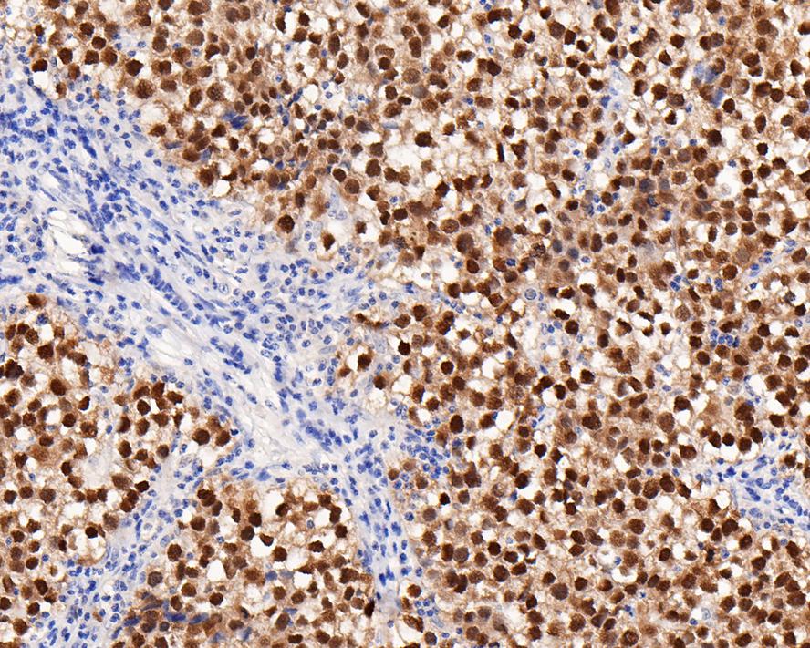
Immunohistochemical analysis of paraffin-embedded human seminoma tissue with Rabbit anti-Oct4 antibody at 1/4,000 dilution. The section was pre-treated using heat mediated antigen retrieval with sodium citrate buffer (pH 6.0) for 2 minutes. The tissues were blocked in 1% BSA for 20 minutes at room temperature, washed with ddH2O and PBS, and then probed with the primary antibody at 1/4,000 dilution for 1 hour at room temperature. The detection was performed using an HRP conjugated compact polymer system. DAB was used as the chromogen. Tissues were counterstained with hematoxylin and mounted with DPX.ICC/IF
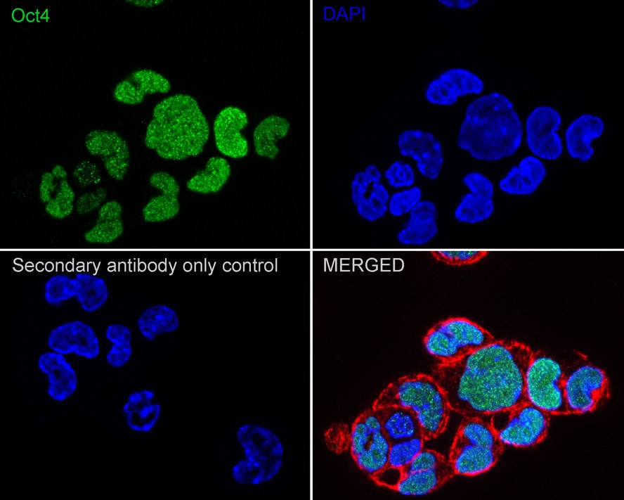
Immunocytochemistry analysis of F9 cells labeling Oct4 with Rabbit anti-Oct4 antibody at 1/200 dilution. Cells were fixed in 4% paraformaldehyde for 20 minutes at room temperature, permeabilized with 0.1% Triton X-100 in PBS for 5 minutes at room temperature, then blocked with 1% BSA in 10% negative goat serum for 1 hour at room temperature. Cells were then incubated with Rabbit anti-Oct4 antibody at 1/200 dilution in 1% BSA in PBST overnight at 4 ℃. Goat Anti-Rabbit IgG H&L (iFluor™ 488) was used as the secondary antibody at 1/1,000 dilution. PBS instead of the primary antibody was used as the secondary antibody only control. Nuclear DNA was labelled in blue with DAPI. Beta tubulin (red) was stained at 1/100 dilution overnight at +4℃. Goat Anti-Mouse IgG H&L (iFluor™ 594) was used as the secondary antibody at 1/1,000 dilution.FC
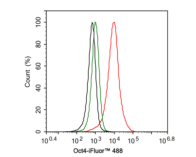
Flow cytometric analysis of NCCIT cells labeling Oct4. Cells were fixed and permeabilized. Then stained with the primary antibody (1ug/ml) (red) compared with Rabbit IgG Isotype Control (green). After incubation of the primary antibody at +4℃ for an hour, the cells were stained with a iFluor™ 488 conjugate-Goat anti-Rabbit IgG Secondary antibody at 1/1,000 dilution for 30 minutes at +4℃. Unlabelled sample was used as a control (cells without incubation with primary antibody; black).IP
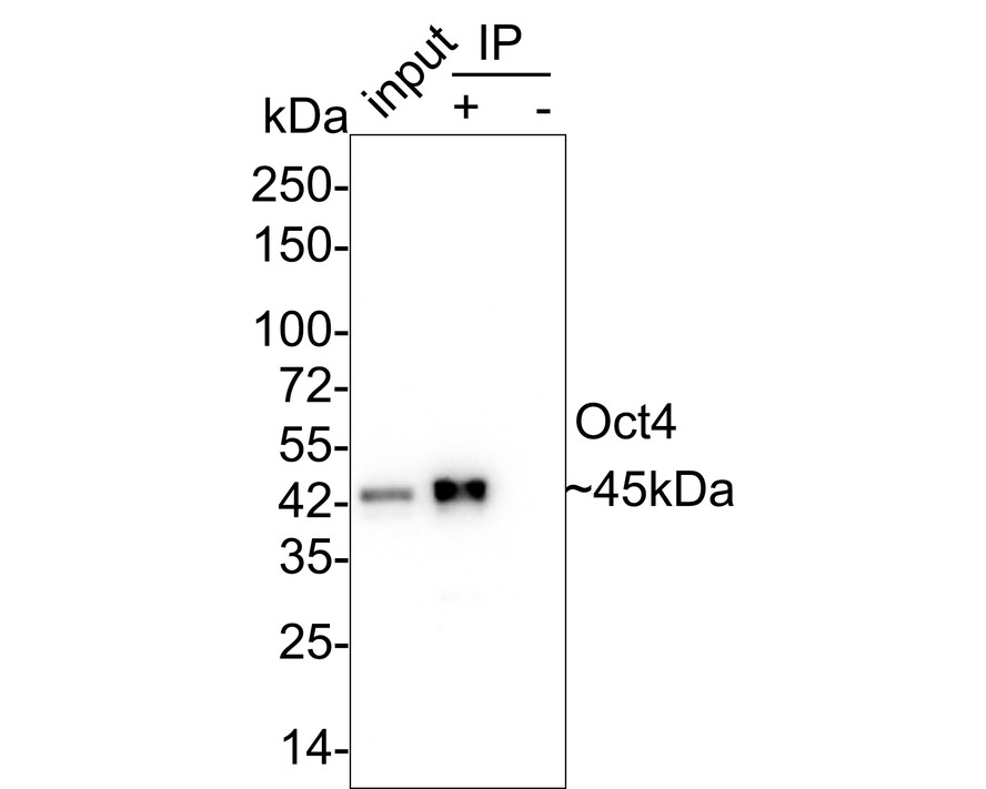
Oct4 was immunoprecipitated in 0.2mg NCCIT cell lysate with Rabbit anti-Oct4 antibody at 2 µg/25 µl agarose. Western blot was performed from the immunoprecipitate using Rabbit anti-Oct4 antibody at 1/2,000 dilution. Anti-Rabbit IgG for IP Nano-secondary antibody at 1/5,000 dilution was used for 1 hour at room temperature. Lane 1: NCCIT cell lysate (input), Lane 2: Rabbit anti-Oct4 antibody IP in NCCIT cell lysate, Lane 3: Rabbit IgG instead of Rabbit anti-Oct4 antibody in NCCIT cell lysate. Blocking/Dilution buffer: 5% NFDM/TBST Exposure time: 1 minute 40 secondsChIP
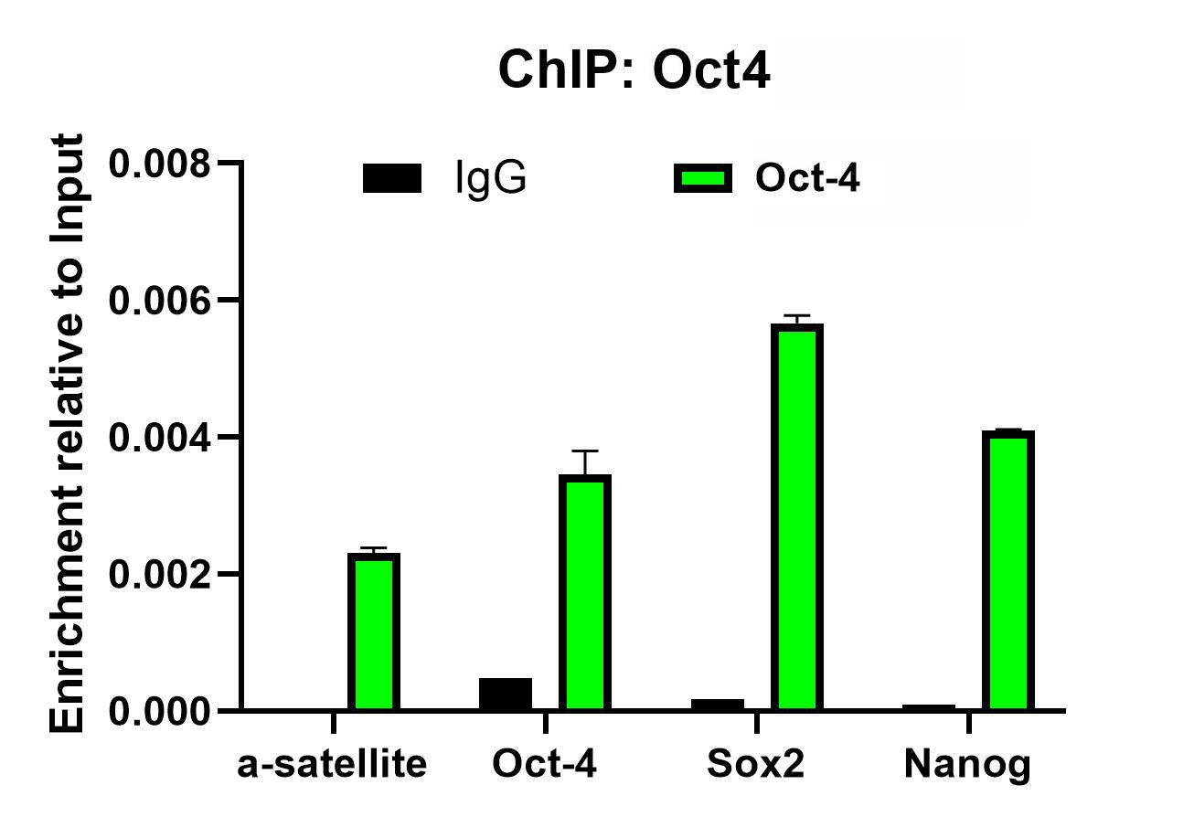
Chromatin immunoprecipitations were performed with cross-linked chromatin from NCCIT cells and either Oct4 or Normal Rabbit IgG according to the ChIP protocol. The enriched DNA was quantified by real-time PCR using indicated primers. The amount of immunoprecipitated DNA in each sample is represented as signal relative to the total amount of input chromatin, which is equivalent to one.| Product Name | Oct4 Recombinant Rabbit Monoclonal Antibody |
|---|---|
| Antibody Type | Primary Antibodies |
| Immunogen | Synthetic peptide within human Oct4 aa 20-60. |
| Clonality | Monoclonal |
|---|---|
| Isotype | IgG |
| Host Species | Rabbit |
| Tested Applications | ChIPFCICC/IFIHCIPWB |
| WB:1:1000-1:8000 IHC:1:500-1:4000 ICC:1:200-1:2000 FC:1ug/ml IP:Use at an assay dependent concentration. ChIP:Use 0.5~2 μg for 25 μg of chromatin. |
|
| Species Reactivity | HumanMouse |
| Concentration | 1mg/ml |
| Purification | Protein A |
| Gene Symbol | POU5F1 |
|---|---|
| Gene Synonyms | OCT3 OCT4 OTF3 OTF4 OTF-3 Oct-3 Oct-4 Oct3/4 |
| Gene Full Name | POU class 5 homeobox 1 |
| Gene Summary | This gene encodes a transcription factor containing a POU homeodomain that plays a key role in embryonic development and stem cell pluripotency. Aberrant expression of this gene in adult tissues is associated with tumorigenesis. This gene can participate in a translocation with the Ewing's sarcoma gene on chromosome 21, which also leads to tumor formation. Alternative splicing, as well as usage of alternative AUG and non-AUG translation initiation codons, results in multiple isoforms. One of the AUG start codons is polymorphic in human populations. Related pseudogenes have been identified on chromosomes 1, 3, 8, 10, and 12. [provided by RefSeq, Oct 2013] |
| Molecular Weight(MW) | 39kDa(Observed band size: 45kDa) |
| Cellular Localization | Cytoplasm, Nucleus. |
WB
Western blot analysis of Oct4 on different lysates with Rabbit anti-Oct4 antibody at 1/1,000 dilution. Lane 1: F9 cell lysate, Lane 2: NCCIT cell lysate, Lysates/proteins at 20 µg/Lane. Exposure time: 1 minute 59 seconds; 4-20% SDS-PAGE gel. Proteins were transferred to a PVDF membrane and blocked with 5% NFDM/TBST for 1 hour at room temperature. The primary antibody at 1/1,000 dilution was used in 5% NFDM/TBST at 4℃ overnight. Goat Anti-Rabbit IgG - HRP Secondary Antibody at 1/50,000 dilution was used for 1 hour at room temperature.
IHC
Immunohistochemical analysis of paraffin-embedded human seminoma tissue with Rabbit anti-Oct4 antibody at 1/4,000 dilution. The section was pre-treated using heat mediated antigen retrieval with sodium citrate buffer (pH 6.0) for 2 minutes. The tissues were blocked in 1% BSA for 20 minutes at room temperature, washed with ddH2O and PBS, and then probed with the primary antibody at 1/4,000 dilution for 1 hour at room temperature. The detection was performed using an HRP conjugated compact polymer system. DAB was used as the chromogen. Tissues were counterstained with hematoxylin and mounted with DPX.
ICC/IF
Immunocytochemistry analysis of F9 cells labeling Oct4 with Rabbit anti-Oct4 antibody at 1/200 dilution. Cells were fixed in 4% paraformaldehyde for 20 minutes at room temperature, permeabilized with 0.1% Triton X-100 in PBS for 5 minutes at room temperature, then blocked with 1% BSA in 10% negative goat serum for 1 hour at room temperature. Cells were then incubated with Rabbit anti-Oct4 antibody at 1/200 dilution in 1% BSA in PBST overnight at 4 ℃. Goat Anti-Rabbit IgG H&L (iFluor™ 488) was used as the secondary antibody at 1/1,000 dilution. PBS instead of the primary antibody was used as the secondary antibody only control. Nuclear DNA was labelled in blue with DAPI. Beta tubulin (red) was stained at 1/100 dilution overnight at +4℃. Goat Anti-Mouse IgG H&L (iFluor™ 594) was used as the secondary antibody at 1/1,000 dilution.
FC
Flow cytometric analysis of NCCIT cells labeling Oct4. Cells were fixed and permeabilized. Then stained with the primary antibody (1ug/ml) (red) compared with Rabbit IgG Isotype Control (green). After incubation of the primary antibody at +4℃ for an hour, the cells were stained with a iFluor™ 488 conjugate-Goat anti-Rabbit IgG Secondary antibody at 1/1,000 dilution for 30 minutes at +4℃. Unlabelled sample was used as a control (cells without incubation with primary antibody; black).
IP
Oct4 was immunoprecipitated in 0.2mg NCCIT cell lysate with Rabbit anti-Oct4 antibody at 2 µg/25 µl agarose. Western blot was performed from the immunoprecipitate using Rabbit anti-Oct4 antibody at 1/2,000 dilution. Anti-Rabbit IgG for IP Nano-secondary antibody at 1/5,000 dilution was used for 1 hour at room temperature. Lane 1: NCCIT cell lysate (input), Lane 2: Rabbit anti-Oct4 antibody IP in NCCIT cell lysate, Lane 3: Rabbit IgG instead of Rabbit anti-Oct4 antibody in NCCIT cell lysate. Blocking/Dilution buffer: 5% NFDM/TBST Exposure time: 1 minute 40 seconds
ChIP
Chromatin immunoprecipitations were performed with cross-linked chromatin from NCCIT cells and either Oct4 or Normal Rabbit IgG according to the ChIP protocol. The enriched DNA was quantified by real-time PCR using indicated primers. The amount of immunoprecipitated DNA in each sample is represented as signal relative to the total amount of input chromatin, which is equivalent to one.| Application Notes | WB:1:1000-1:8000 IHC:1:500-1:4000 ICC:1:200-1:2000 FC:1ug/ml IP:Use at an assay dependent concentration. ChIP:Use 0.5~2 μg for 25 μg of chromatin. |
|---|
| Form | Liquid |
|---|---|
| Storage Instructions | Store at +4℃ after thawing. Aliquot store at -20℃ or -80℃. Avoid repeated freeze / thaw cycles. |
| Storage Buffer | 1*TBS (pH7.4), 0.05% BSA, 40% Glycerol. Preservative: 0.05% Sodium Azide. |


 Data sheet for OM643256