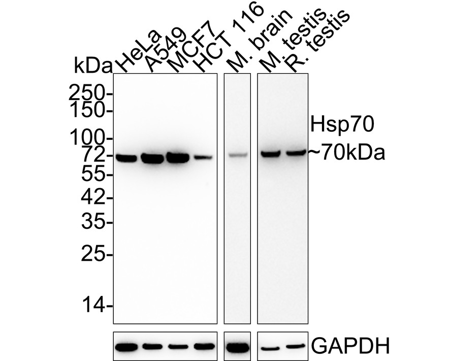
WB
Western blot analysis of Hsp70 on different lysates with Rabbit anti-Hsp70 antibody at 1/2,000 dilution. Lane 1: HeLa cell lysate, Lane 2: A549 cell lysate, Lane 3: MCF7 cell lysate, Lane 4: HCT 116 cell lysate, Lane 5: Mouse brain tissue lysate, Lane 6: Mouse testis tissue lysate, Lane 7: Rat testis tissue lysate, Lysates/proteins at 15 µg/Lane. Exposure time: Lane 1-4: 43 seconds; Lane 5-7: 2 minutes; 4-20% SDS-PAGE gel. Proteins were transferred to a PVDF membrane and blocked with 5% NFDM/TBST for 1 hour at room temperature. The primary antibody at 1/2,000 dilution was used in 5% NFDM/TBST at 4℃ overnight. Goat Anti-Rabbit IgG - HRP Secondary Antibody at 1:50,000 dilution was used for 1 hour at room temperature.IHC
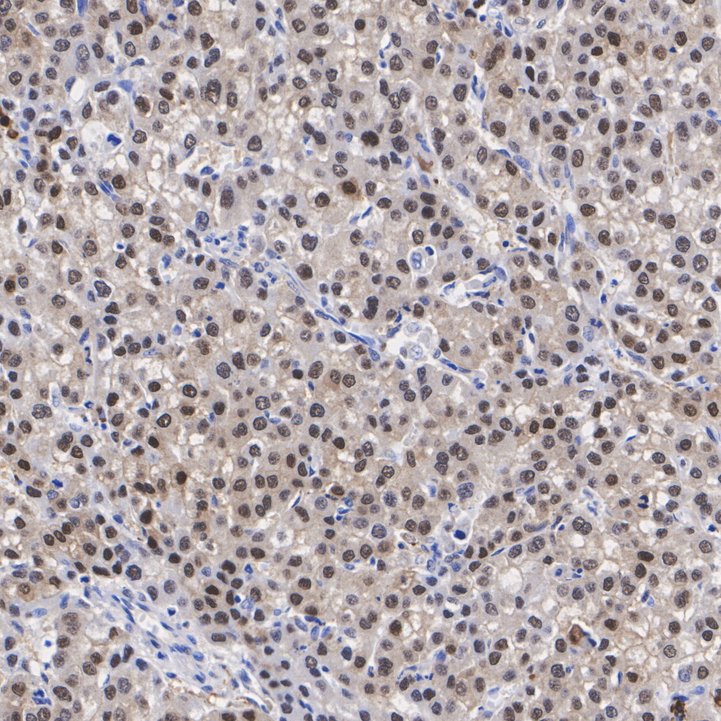
Immunohistochemical analysis of paraffin-embedded human liver cancer tissue with Rabbit anti-Hsp70 antibody at 1/500 dilution. The section was pre-treated using heat mediated antigen retrieval with Tris-EDTA buffer (pH 9.0) for 20 minutes. The tissues were blocked in 1% BSA for 20 minutes at room temperature, washed with ddH2O and PBS, and then probed with the primary antibody at 1/500 dilution for 1 hour at room temperature. The detection was performed using an HRP conjugated compact polymer system. DAB was used as the chromogen. Tissues were counterstained with hematoxylin and mounted with DPX.ICC/IF
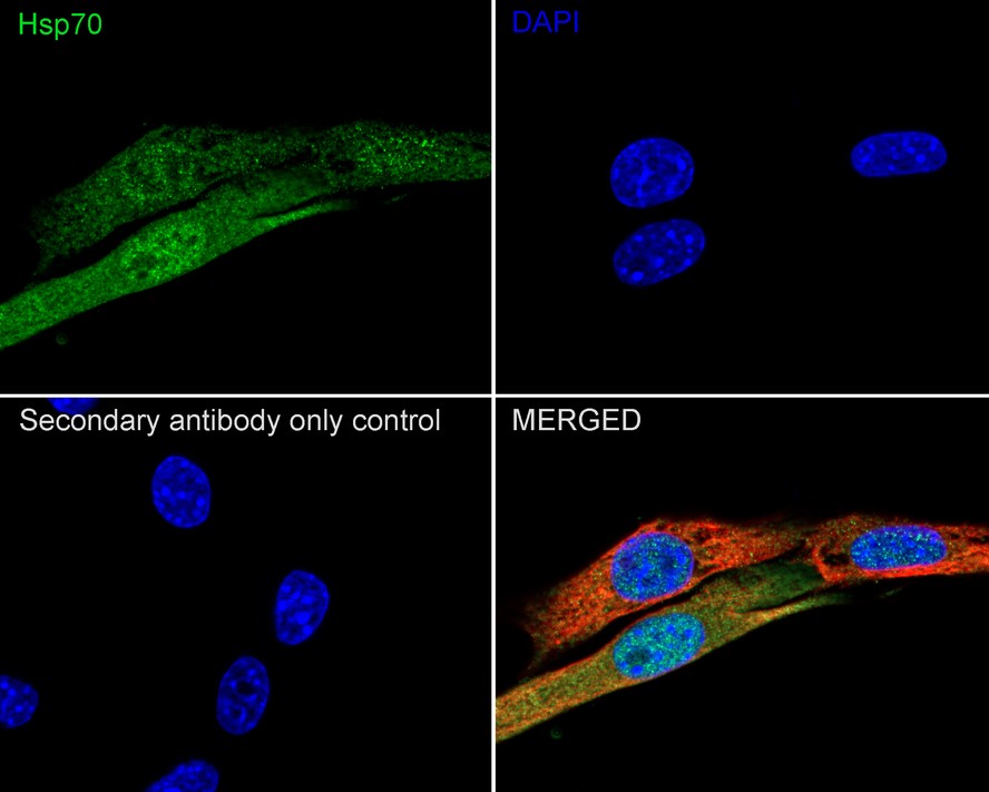
Immunocytochemistry analysis of NIH/3T3 cells labeling Hsp70 with Rabbit anti-Hsp70 antibody at 1/100 dilution. Cells were fixed in 4% paraformaldehyde for 20 minutes at room temperature, permeabilized with 0.1% Triton X-100 in PBS for 5 minutes at room temperature, then blocked with 1% BSA in 10% negative goat serum for 1 hour at room temperature. Cells were then incubated with Rabbit anti-Hsp70 antibody at 1/100 dilution in 1% BSA in PBST overnight at 4 ℃. Goat Anti-Rabbit IgG H&L (iFluor™ 488) was used as the secondary antibody at 1/1,000 dilution. PBS instead of the primary antibody was used as the secondary antibody only control. Nuclear DNA was labelled in blue with DAPI. Beta tubulin (red) was stained at 1/100 dilution overnight at +4℃. Goat Anti-Mouse IgG H&L (iFluor™ 594) was used as the secondary antibody at 1/1,000 dilution.FC
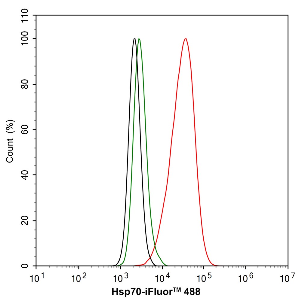
Flow cytometric analysis of A549 cells labeling Hsp70. Cells were fixed and permeabilized. Then stained with the primary antibody (1/100) (red) compared with Rabbit IgG Isotype Control (green). After incubation of the primary antibody at +4℃ for an hour, the cells were stained with a iFluor™ 488 conjugate-Goat anti-Rabbit IgG Secondary antibody at 1/1,000 dilution for 30 minutes at +4℃. Unlabelled sample was used as a control (cells without incubation with primary antibody; black).| Product Name | Hsp70 Recombinant Rabbit Monoclonal Antibody |
|---|---|
| Antibody Type | Primary Antibodies |
| Immunogen | Recombinant protein within Mouse Hsp70 aa 403-641 / 641. |
| Clonality | monoclonal |
|---|---|
| Isotype | IgG |
| Host Species | Rabbit |
| Tested Applications | FCICC/IFIHCWB |
| WB:1:2000-1:5000 IHC:1:500-1:1500 ICC:1:100 FC:1:100 |
|
| Species Reactivity | HumanMouseRat |
| Concentration | 1mg/ml |
| Purification | Protein A |
| Gene Symbol | Hspa1a |
|---|---|
| Gene Synonyms | HSP70 HSP72 HSPA1 HSP70I HSP70-1 HSP70-2 HSP70.1 HSP70.2 HSP70-1A HEL-S-103 |
| Gene Full Name | heat shock protein family A (Hsp70) member 1A |
| Gene Summary | This intronless gene encodes a 70kDa heat shock protein which is a member of the heat shock protein 70 family. In conjuction with other heat shock proteins, this protein stabilizes existing proteins against aggregation and mediates the folding of newly translated proteins in the cytosol and in organelles. It is also involved in the ubiquitin-proteasome pathway through interaction with the AU-rich element RNA-binding protein 1. The gene is located in the major histocompatibility complex class III region, in a cluster with two closely related genes which encode similar proteins. [provided by RefSeq, Jul 2008] |
| Molecular Weight(MW) | 70kDa |
| Cellular Localization | Cytoplasm. |
WB
Western blot analysis of Hsp70 on different lysates with Rabbit anti-Hsp70 antibody at 1/2,000 dilution. Lane 1: HeLa cell lysate, Lane 2: A549 cell lysate, Lane 3: MCF7 cell lysate, Lane 4: HCT 116 cell lysate, Lane 5: Mouse brain tissue lysate, Lane 6: Mouse testis tissue lysate, Lane 7: Rat testis tissue lysate, Lysates/proteins at 15 µg/Lane. Exposure time: Lane 1-4: 43 seconds; Lane 5-7: 2 minutes; 4-20% SDS-PAGE gel. Proteins were transferred to a PVDF membrane and blocked with 5% NFDM/TBST for 1 hour at room temperature. The primary antibody at 1/2,000 dilution was used in 5% NFDM/TBST at 4℃ overnight. Goat Anti-Rabbit IgG - HRP Secondary Antibody at 1:50,000 dilution was used for 1 hour at room temperature.
IHC
Immunohistochemical analysis of paraffin-embedded human liver cancer tissue with Rabbit anti-Hsp70 antibody at 1/500 dilution. The section was pre-treated using heat mediated antigen retrieval with Tris-EDTA buffer (pH 9.0) for 20 minutes. The tissues were blocked in 1% BSA for 20 minutes at room temperature, washed with ddH2O and PBS, and then probed with the primary antibody at 1/500 dilution for 1 hour at room temperature. The detection was performed using an HRP conjugated compact polymer system. DAB was used as the chromogen. Tissues were counterstained with hematoxylin and mounted with DPX.
ICC/IF
Immunocytochemistry analysis of NIH/3T3 cells labeling Hsp70 with Rabbit anti-Hsp70 antibody at 1/100 dilution. Cells were fixed in 4% paraformaldehyde for 20 minutes at room temperature, permeabilized with 0.1% Triton X-100 in PBS for 5 minutes at room temperature, then blocked with 1% BSA in 10% negative goat serum for 1 hour at room temperature. Cells were then incubated with Rabbit anti-Hsp70 antibody at 1/100 dilution in 1% BSA in PBST overnight at 4 ℃. Goat Anti-Rabbit IgG H&L (iFluor™ 488) was used as the secondary antibody at 1/1,000 dilution. PBS instead of the primary antibody was used as the secondary antibody only control. Nuclear DNA was labelled in blue with DAPI. Beta tubulin (red) was stained at 1/100 dilution overnight at +4℃. Goat Anti-Mouse IgG H&L (iFluor™ 594) was used as the secondary antibody at 1/1,000 dilution.
FC
Flow cytometric analysis of A549 cells labeling Hsp70. Cells were fixed and permeabilized. Then stained with the primary antibody (1/100) (red) compared with Rabbit IgG Isotype Control (green). After incubation of the primary antibody at +4℃ for an hour, the cells were stained with a iFluor™ 488 conjugate-Goat anti-Rabbit IgG Secondary antibody at 1/1,000 dilution for 30 minutes at +4℃. Unlabelled sample was used as a control (cells without incubation with primary antibody; black).| Application Notes | WB:1:2000-1:5000 IHC:1:500-1:1500 ICC:1:100 FC:1:100 |
|---|
| Form | Liquid |
|---|---|
| Storage Instructions | Store at +4℃ after thawing. Aliquot store at -20℃ or -80℃. Avoid repeated freeze / thaw cycles. |
| Storage Buffer | 1*TBS (pH7.4), 0.05% BSA, 40% Glycerol. Preservative: 0.05% Sodium Azide. |


 Data sheet for OM643861