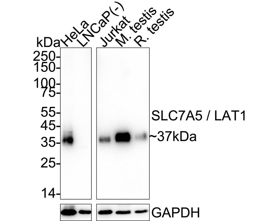
WB
Western blot analysis of SLC7A5 / LAT1 on different lysates with Rabbit anti-SLC7A5 / LAT1 antibody at 1/2,000 dilution. Lane 1: HeLa cell lysate (20 µg/Lane), Lane 2: LNCaP cell lysate (negative) (20 µg/Lane), Lane 3: Jurkat cell lysate (20 µg/Lane), Lane 4: Mouse testis tissue lysate (40 µg/Lane), Lane 5: Rat testis tissue lysate (40 µg/Lane), Exposure time: Lane 1-2: 1 minute; Lane 3-5: 3 minutes; 4-20% SDS-PAGE gel. Proteins were transferred to a PVDF membrane and blocked with 5% NFDM/TBST for 1 hour at room temperature. The primary antibody at 1/2,000 dilution was used in 5% NFDM/TBST at 4℃ overnight. Goat Anti-Rabbit IgG - HRP Secondary Antibody at 1/50,000 dilution was used for 1 hour at room temperature.IHC
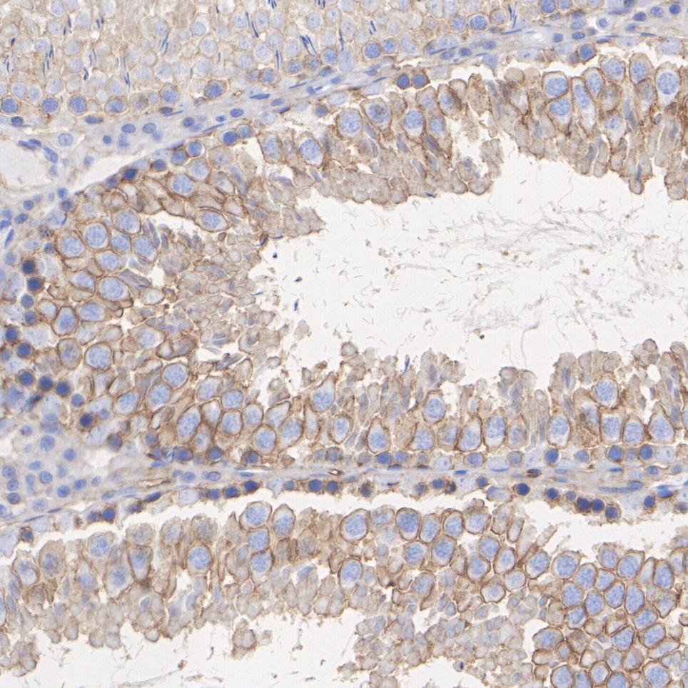
Immunohistochemical analysis of paraffin-embedded rat testis tissue with Rabbit anti-SLC7A5 / LAT1 antibody at 1/200 dilution. The section was pre-treated using heat mediated antigen retrieval with Tris-EDTA buffer (pH 9.0) for 20 minutes. The tissues were blocked in 1% BSA for 20 minutes at room temperature, washed with ddH2O and PBS, and then probed with the primary antibody at 1/200 dilution for 1 hour at room temperature. The detection was performed using an HRP conjugated compact polymer system. DAB was used as the chromogen. Tissues were counterstained with hematoxylin and mounted with DPX.ICC/IF
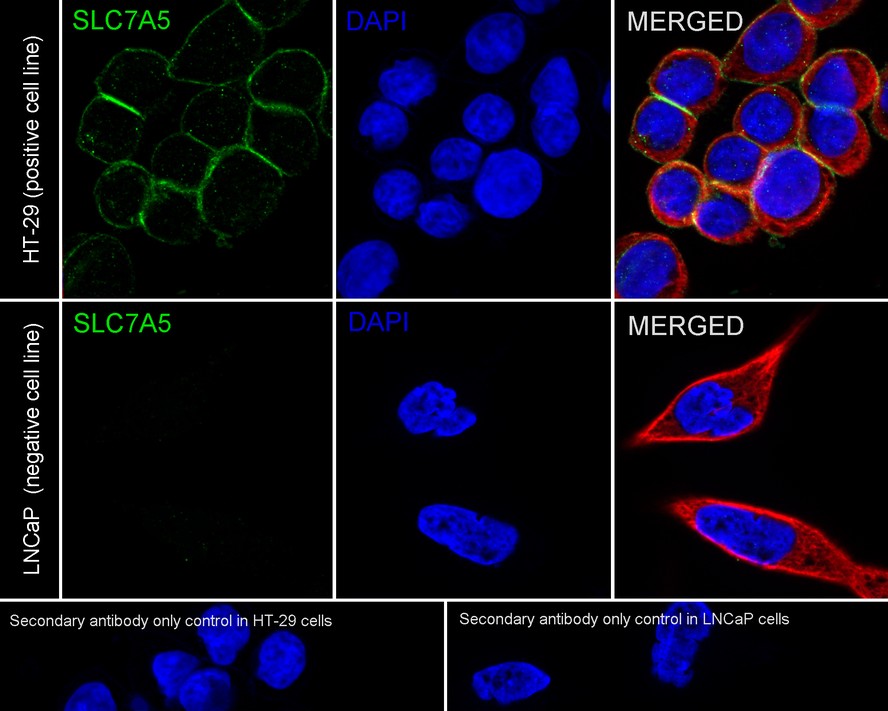
Immunocytochemistry analysis of HT-29 (positive) and LNCaP (negative) labeling SLC7A5 / LAT1 with Rabbit anti-SLC7A5 / LAT1 antibody at 1/100 dilution. Cells were fixed in 4% paraformaldehyde for 15 minutes at room temperature, permeabilized with 0.1% Triton X-100 in PBS for 15 minutes at room temperature, then blocked with 1% BSA in 10% negative goat serum for 1 hour at room temperature. Cells were then incubated with Rabbit anti-SLC7A5 / LAT1 antibody at 1/100 dilution in 1% BSA in PBST overnight at 4 ℃. Goat Anti-Rabbit IgG H&L (iFluor™ 488) was used as the secondary antibody at 1/1,000 dilution. PBS instead of the primary antibody was used as the secondary antibody only control. Nuclear DNA was labelled in blue with DAPI. Beta tubulin (red) was stained at 1/100 dilution overnight at +4℃. Goat Anti-Mouse IgG H&L (iFluor™ 594) was used as the secondary antibody at 1/1,000 dilution.IP
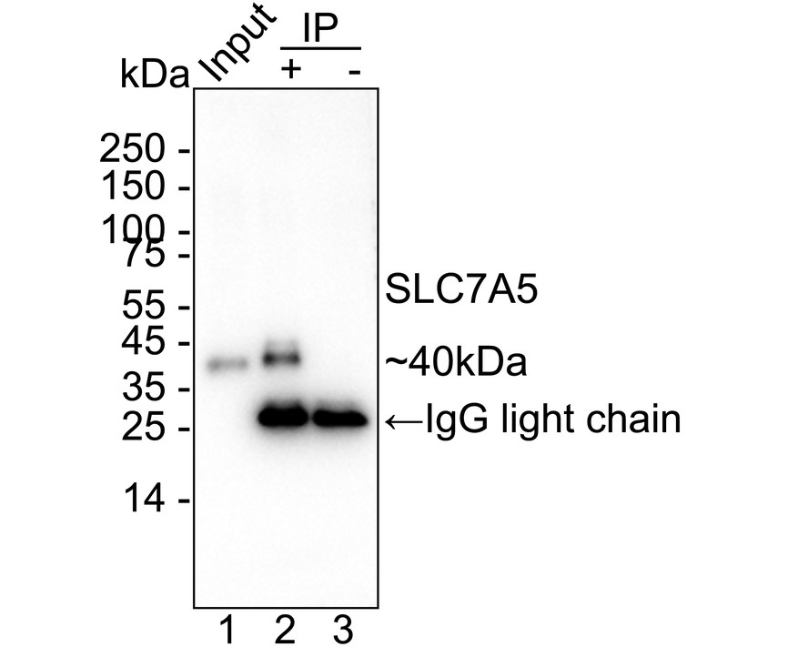
SLC7A5 / LAT1 was immunoprecipitated from 0.2 mg A549 cell lysate with Rabbit anti-SLC7A5 / LAT1 antibody at 2 µg/25 µl agarose. Western blot was performed from the immunoprecipitate using Rabbit anti-SLC7A5 / LAT1 antibody at 1/1,000 dilution. Mouse Anti-Rabbit IgG kappa light chain secondary antibody at 1/5,000 dilution was used for 1 hour at room temperature. Lane 1: A549 cell lysate (input), Lane 2: Rabbit anti-SLC7A5 / LAT1 antibody IP in A549 cell lysate, Lane 3: Rabbit IgG instead of Rabbit anti-SLC7A5 / LAT1 antibody in A549 cell lysate. Blocking/Dilution buffer: 5% NFDM/TBST. Exposure time: 59 seconds.| Product Name | SLC7A5 / LAT1 Recombinant Rabbit Monoclonal Antibody |
|---|---|
| Antibody Type | Primary Antibodies |
| Immunogen | Recombinant protein within human SLC7A5 aa 71-507. |
| Clonality | monoclonal |
|---|---|
| Isotype | IgG |
| Host Species | Rabbit |
| Tested Applications | ICC/IFIHCIPWB |
| WB:1:2000 IHC:1:200 ICC/IF:1:100 IP:1-2μg/sample |
|
| Species Reactivity | HumanMouseRat |
| Concentration | 1mg/ml |
| Purification | Protein A |
| Gene Symbol | SLC7A5 |
|---|---|
| Gene Synonyms | E16 CD98 LAT1 4F2LC MPE16 D16S469E |
| Gene Full Name | solute carrier family 7 member 5 |
| Gene Summary | Enables L-amino acid transmembrane transporter activity and secondary active transmembrane transporter activity. Involved in carboxylic acid transport; thyroid hormone transport; and xenobiotic transport. Located in several cellular components, including apical plasma membrane; cytosol; and microvillus membrane. Part of amino acid transport complex. Implicated in cholangiocarcinoma; colon cancer; hepatocellular carcinoma; and lung squamous cell carcinoma. Biomarker of esophagitis; gastrointestinal system cancer (multiple); malignant astrocytoma (multiple); and respiratory system cancer (multiple). [provided by Alliance of Genome Resources, Feb 2025] |
| Molecular Weight(MW) | 55kDa(Observed band size: 37kDa) |
| Cellular Localization | Apical cell membrane, Cell membrane, Lysosome membrane. |
WB
Western blot analysis of SLC7A5 / LAT1 on different lysates with Rabbit anti-SLC7A5 / LAT1 antibody at 1/2,000 dilution. Lane 1: HeLa cell lysate (20 µg/Lane), Lane 2: LNCaP cell lysate (negative) (20 µg/Lane), Lane 3: Jurkat cell lysate (20 µg/Lane), Lane 4: Mouse testis tissue lysate (40 µg/Lane), Lane 5: Rat testis tissue lysate (40 µg/Lane), Exposure time: Lane 1-2: 1 minute; Lane 3-5: 3 minutes; 4-20% SDS-PAGE gel. Proteins were transferred to a PVDF membrane and blocked with 5% NFDM/TBST for 1 hour at room temperature. The primary antibody at 1/2,000 dilution was used in 5% NFDM/TBST at 4℃ overnight. Goat Anti-Rabbit IgG - HRP Secondary Antibody at 1/50,000 dilution was used for 1 hour at room temperature.
IHC
Immunohistochemical analysis of paraffin-embedded rat testis tissue with Rabbit anti-SLC7A5 / LAT1 antibody at 1/200 dilution. The section was pre-treated using heat mediated antigen retrieval with Tris-EDTA buffer (pH 9.0) for 20 minutes. The tissues were blocked in 1% BSA for 20 minutes at room temperature, washed with ddH2O and PBS, and then probed with the primary antibody at 1/200 dilution for 1 hour at room temperature. The detection was performed using an HRP conjugated compact polymer system. DAB was used as the chromogen. Tissues were counterstained with hematoxylin and mounted with DPX.
ICC/IF
Immunocytochemistry analysis of HT-29 (positive) and LNCaP (negative) labeling SLC7A5 / LAT1 with Rabbit anti-SLC7A5 / LAT1 antibody at 1/100 dilution. Cells were fixed in 4% paraformaldehyde for 15 minutes at room temperature, permeabilized with 0.1% Triton X-100 in PBS for 15 minutes at room temperature, then blocked with 1% BSA in 10% negative goat serum for 1 hour at room temperature. Cells were then incubated with Rabbit anti-SLC7A5 / LAT1 antibody at 1/100 dilution in 1% BSA in PBST overnight at 4 ℃. Goat Anti-Rabbit IgG H&L (iFluor™ 488) was used as the secondary antibody at 1/1,000 dilution. PBS instead of the primary antibody was used as the secondary antibody only control. Nuclear DNA was labelled in blue with DAPI. Beta tubulin (red) was stained at 1/100 dilution overnight at +4℃. Goat Anti-Mouse IgG H&L (iFluor™ 594) was used as the secondary antibody at 1/1,000 dilution.
IP
SLC7A5 / LAT1 was immunoprecipitated from 0.2 mg A549 cell lysate with Rabbit anti-SLC7A5 / LAT1 antibody at 2 µg/25 µl agarose. Western blot was performed from the immunoprecipitate using Rabbit anti-SLC7A5 / LAT1 antibody at 1/1,000 dilution. Mouse Anti-Rabbit IgG kappa light chain secondary antibody at 1/5,000 dilution was used for 1 hour at room temperature. Lane 1: A549 cell lysate (input), Lane 2: Rabbit anti-SLC7A5 / LAT1 antibody IP in A549 cell lysate, Lane 3: Rabbit IgG instead of Rabbit anti-SLC7A5 / LAT1 antibody in A549 cell lysate. Blocking/Dilution buffer: 5% NFDM/TBST. Exposure time: 59 seconds.| Application Notes | WB:1:2000 IHC:1:200 ICC/IF:1:100 IP:1-2μg/sample |
|---|
| Form | Liquid |
|---|---|
| Storage Instructions | Store at +4℃ after thawing. Aliquot store at -20℃ or -80℃. Avoid repeated freeze / thaw cycles. |
| Storage Buffer | 1*TBS (pH7.4), 0.05% BSA, 40% Glycerol. Preservative: 0.05% Sodium Azide. |


 Data sheet for OM643911