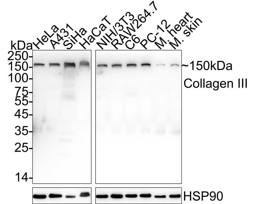
WB
Western blot analysis of Collagen III on different lysates with Rabbit anti-Collagen III antibody at 1/1,000 dilution. Lane 1: HeLa cell lysate (20 µg/Lane) Lane 2: A431 cell lysate (20 µg/Lane) Lane 3: SiHa cell lysate (20 µg/Lane) Lane 4: HaCaT cell lysate (20 µg/Lane) Lane 5: NIH/3T3 cell lysate (20 µg/Lane) Lane 6: RAW264.7 cell lysate (20 µg/Lane) Lane 7: C6 cell lysate (20 µg/Lane) Lane 8: PC-12 cell lysate (20 µg/Lane) Lane 9: Mouse heart tissue lysate (20 µg/Lane) Lane 10: Mouse skin tissue lysate (20 µg/Lane) Exposure time: 2 seconds; 4-20% SDS-PAGE gel. Proteins were transferred to a PVDF membrane and blocked with 5% NFDM/TBST for 1 hour at room temperature. The primary antibody at 1/1,000 dilution was used in 5% NFDM/TBST at 4℃ overnight. Goat Anti-Rabbit IgG - HRP Secondary Antibody at 1/50,000 dilution was used for 1 hour at room temperature.ICC/IF
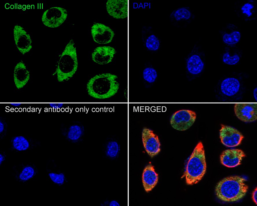
Immunocytochemistry analysis of SiHa cells labeling Collagen III with Rabbit anti-Collagen III antibody at 1/500 dilution. Cells were fixed in 4% paraformaldehyde for 15 minutes at room temperature, permeabilized with 0.1% Triton X-100 in PBS for 15 minutes at room temperature, then blocked with 1% BSA in 10% negative goat serum for 1 hour at room temperature. Cells were then incubated with Rabbit anti-Collagen III antibody at 1/500 dilution in 1% BSA in PBST overnight at 4 ℃. Goat Anti-Rabbit IgG H&L (488) was used as the secondary antibody at 1/1,000 dilution. PBS instead of the primary antibody was used as the secondary antibody only control. Nuclear DNA was labelled in blue with DAPI. Beta tubulin (red) was stained at 1/100 dilution overnight at +4℃. Goat Anti-Mouse IgG H&L (iFluor™ 594) was used as the secondary antibody at 1/1,000 dilution.FC
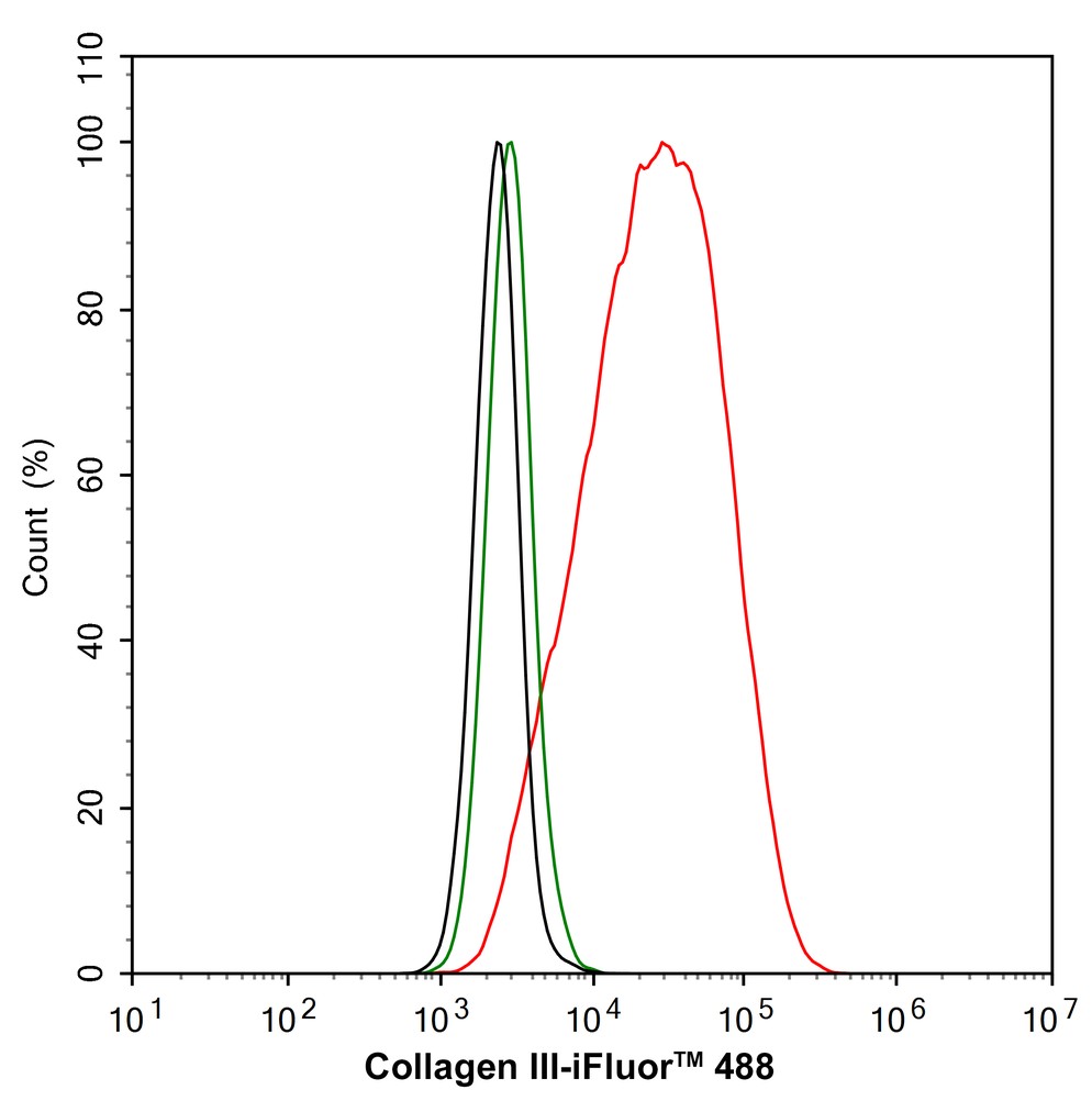
Flow cytometric analysis of HeLa cells labeling Collagen III. Cells were fixed and permeabilized. Then stained with the primary antibody (1/1,000) (red) compared with Rabbit IgG Isotype Control (green). After incubation of the primary antibody at +4℃ for an hour, the cells were stained with a iFluor™ 488 conjugate-Goat anti-Rabbit IgG Secondary antibody at 1/1,000 dilution for 30 minutes at +4℃. Unlabelled sample was used as a control (cells without incubation with primary antibody; black).| Product Name | Collagen III Recombinant Rabbit Monoclonal Antibody |
|---|---|
| Antibody Type | Primary Antibodies |
| Immunogen | Recombinant protein within human Collagen III 101-400/1,466. |
| Clonality | monoclonal |
|---|---|
| Isotype | IgG |
| Host Species | Rabbit |
| Tested Applications | FCICC/IFWB |
| WB:1:500-1:2000 ICC/IF:1:100-1:500 FC:1:500-1:1000 |
|
| Species Reactivity | HumanMouseRat |
| Concentration | 1mg/ml |
| Purification | Protein A |
| Gene Symbol | COL3A1 |
|---|---|
| Gene Synonyms | EDS4A EDSVASC PMGEDSV |
| Gene Full Name | collagen type III alpha 1 chain |
| Gene Summary | This gene encodes the pro-alpha1 chains of type III collagen, a fibrillar collagen that is found in extensible connective tissues such as skin, lung, uterus, intestine and the vascular system, frequently in association with type I collagen. Mutations in this gene are associated with Ehlers-Danlos syndrome type IV, and with aortic and arterial aneurysms. [provided by R. Dalgleish, Feb 2008] |
| Molecular Weight(MW) | 139kDa(Observed band size: 150kDa) |
| Cellular Localization | Extracellular matrix. |
WB
Western blot analysis of Collagen III on different lysates with Rabbit anti-Collagen III antibody at 1/1,000 dilution. Lane 1: HeLa cell lysate (20 µg/Lane) Lane 2: A431 cell lysate (20 µg/Lane) Lane 3: SiHa cell lysate (20 µg/Lane) Lane 4: HaCaT cell lysate (20 µg/Lane) Lane 5: NIH/3T3 cell lysate (20 µg/Lane) Lane 6: RAW264.7 cell lysate (20 µg/Lane) Lane 7: C6 cell lysate (20 µg/Lane) Lane 8: PC-12 cell lysate (20 µg/Lane) Lane 9: Mouse heart tissue lysate (20 µg/Lane) Lane 10: Mouse skin tissue lysate (20 µg/Lane) Exposure time: 2 seconds; 4-20% SDS-PAGE gel. Proteins were transferred to a PVDF membrane and blocked with 5% NFDM/TBST for 1 hour at room temperature. The primary antibody at 1/1,000 dilution was used in 5% NFDM/TBST at 4℃ overnight. Goat Anti-Rabbit IgG - HRP Secondary Antibody at 1/50,000 dilution was used for 1 hour at room temperature.
ICC/IF
Immunocytochemistry analysis of SiHa cells labeling Collagen III with Rabbit anti-Collagen III antibody at 1/500 dilution. Cells were fixed in 4% paraformaldehyde for 15 minutes at room temperature, permeabilized with 0.1% Triton X-100 in PBS for 15 minutes at room temperature, then blocked with 1% BSA in 10% negative goat serum for 1 hour at room temperature. Cells were then incubated with Rabbit anti-Collagen III antibody at 1/500 dilution in 1% BSA in PBST overnight at 4 ℃. Goat Anti-Rabbit IgG H&L (488) was used as the secondary antibody at 1/1,000 dilution. PBS instead of the primary antibody was used as the secondary antibody only control. Nuclear DNA was labelled in blue with DAPI. Beta tubulin (red) was stained at 1/100 dilution overnight at +4℃. Goat Anti-Mouse IgG H&L (iFluor™ 594) was used as the secondary antibody at 1/1,000 dilution.
FC
Flow cytometric analysis of HeLa cells labeling Collagen III. Cells were fixed and permeabilized. Then stained with the primary antibody (1/1,000) (red) compared with Rabbit IgG Isotype Control (green). After incubation of the primary antibody at +4℃ for an hour, the cells were stained with a iFluor™ 488 conjugate-Goat anti-Rabbit IgG Secondary antibody at 1/1,000 dilution for 30 minutes at +4℃. Unlabelled sample was used as a control (cells without incubation with primary antibody; black).| Application Notes | WB:1:500-1:2000 ICC/IF:1:100-1:500 FC:1:500-1:1000 |
|---|
| Form | Liquid |
|---|---|
| Storage Instructions | Store at +4℃ after thawing. Aliquot store at -20℃. Avoid repeated freeze / thaw cycles. |
| Storage Buffer | 1*TBS (pH7.4), 0.05% BSA, 40% Glycerol. Preservative: 0.05% Sodium Azide. |


 Data sheet for OM644016