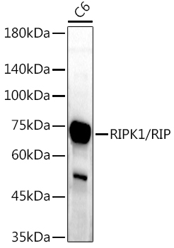
WB
Western blot analysis of lysates from C6 cells, using RIPK1/RIP Rabbit pAb at 1:1000 dilution. Secondary antibody: HRP Goat Anti-Rabbit IgG (H+L) at 1:10000 dilution. Lysates/proteins: 25μg per lane. Blocking buffer: 3% nonfat dry milk in TBST. Detection: ECL Enhanced Kit. Exposure time: 180s.IHC
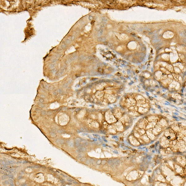
Immunohistochemistry analysis of paraffin embedded Mouse colon using RIPK1/RIP Rabbit pAb at dilution of 1:50 (40x lens). High pressure antigen retrieval performed with 0.01M Citrate Bufferr (pH 6.0) prior to IHC staining.ICC/IF
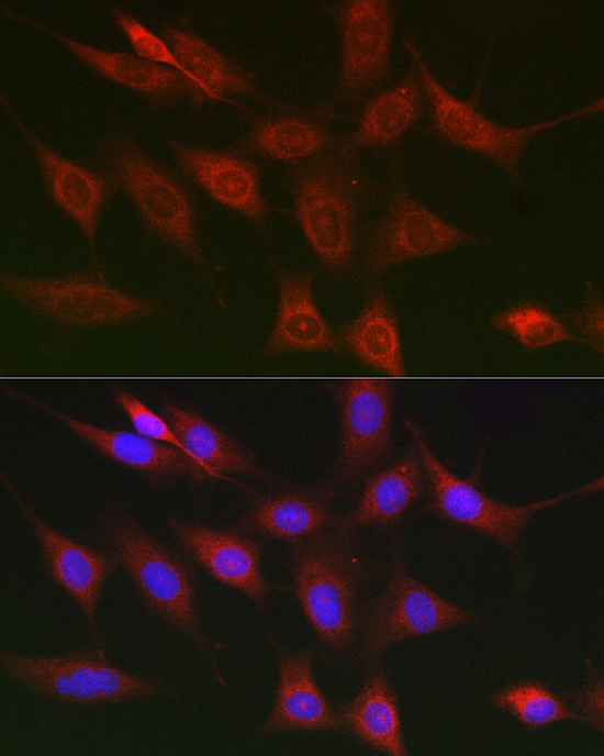
Immunofluorescence analysis of NIH/3T3 cells using RIPK1/RIP Rabbit pAb at dilution of 1:100 (40x lens). Secondary antibody: Cy3 Goat Anti-Rabbit IgG (H+L) at 1:500 dilution. Blue: DAPI for nuclear staining.| Product Name | RIPK1/RIP Rabbit pAb |
|---|---|
| Antibody Type | Primary Antibodies |
| Immunogen | A synthetic peptide corresponding to a sequence within amino acids 100-200 of human RIPK1/RIP (NP_003795.2). |
| Clonality | polyclonal |
|---|---|
| Isotype | IgG |
| Host Species | Rabbit |
| Tested Applications | ICC/IFIHCWB |
| WB:1:500-1:1000 IHC:1:50-1:200 ICC/IF:1:50-1:200 |
|
| Species Reactivity | HumanMouseRat |
| Concentration | 1mg/ml |
| Purification | Affinity purified |
| Gene Symbol | RIPK1 |
|---|---|
| Gene Synonyms | RIP RIP1 AIEFL IMD57 RIP-1 |
| Gene Full Name | receptor interacting serine/threonine kinase 1 |
| Gene Summary | This gene encodes a member of the receptor-interacting protein (RIP) family of serine/threonine protein kinases. The encoded protein plays a role in inflammation and cell death in response to tissue damage, pathogen recognition, and as part of developmental regulation. RIPK1/RIPK3 kinase-mediated necrosis is referred to as necroptosis. Genetic disruption of this gene in mice results in death shortly after birth. [provided by RefSeq, Aug 2017] |
| Molecular Weight(MW) | 76kDa(Observed MW 75/38kDa) |
| Cellular Localization | Cell membrane, Cytoplasm. |
WB
Western blot analysis of lysates from C6 cells, using RIPK1/RIP Rabbit pAb at 1:1000 dilution. Secondary antibody: HRP Goat Anti-Rabbit IgG (H+L) at 1:10000 dilution. Lysates/proteins: 25μg per lane. Blocking buffer: 3% nonfat dry milk in TBST. Detection: ECL Enhanced Kit. Exposure time: 180s.
IHC
Immunohistochemistry analysis of paraffin embedded Mouse colon using RIPK1/RIP Rabbit pAb at dilution of 1:50 (40x lens). High pressure antigen retrieval performed with 0.01M Citrate Bufferr (pH 6.0) prior to IHC staining.
ICC/IF
Immunofluorescence analysis of NIH/3T3 cells using RIPK1/RIP Rabbit pAb at dilution of 1:100 (40x lens). Secondary antibody: Cy3 Goat Anti-Rabbit IgG (H+L) at 1:500 dilution. Blue: DAPI for nuclear staining.| Application Notes | WB:1:500-1:1000 IHC:1:50-1:200 ICC/IF:1:50-1:200 |
|---|
| Form | Liquid |
|---|---|
| Storage Instructions | Store at -20℃. Avoid freeze / thaw cycles. |
| Storage Buffer | Buffer: PBS with 0.01% thimerosal, 50% glycerol, pH7.3. |


 Data sheet for OM644080