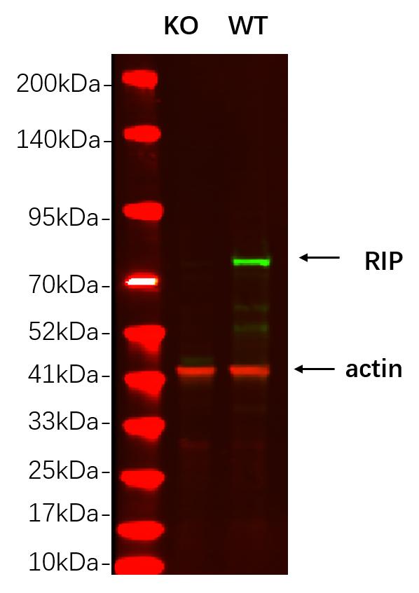
WB
Western blot analysis of lysates from HAP1 WT and knockout cell , (Green) primary antibody was diluted at 1:5000, 4°over night, Dylight 800 secondary antibody was diluted at 1:10000, 37° 1hour. (Red) GAPDH Monoclonal Antibody was diluted at 1:5000 as loading control, 4° over night, Dylight 680 secondary antibody was diluted at 1:10000, 37° 1hour.WB
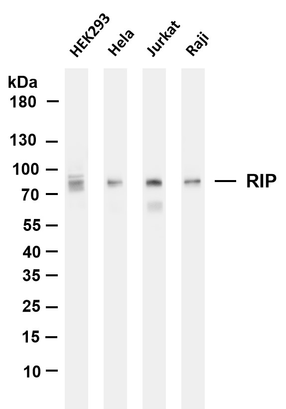
Various whole cell lysates were separated by 4-20% SDS-PAGE, and the membrane was blotted with anti-RIP antibody. The HRP-conjugated Goat anti-Rabbit IgG(H + L) antibody was used to detect the antibody. Lane 1: HEK293, Lane 2: Hela, Lane 3: Jurkat, Lane 4: Raji.IHC
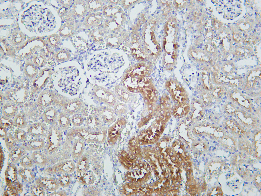
Rat kidney was stained with Anti-RIP rabbit antibody.IHC
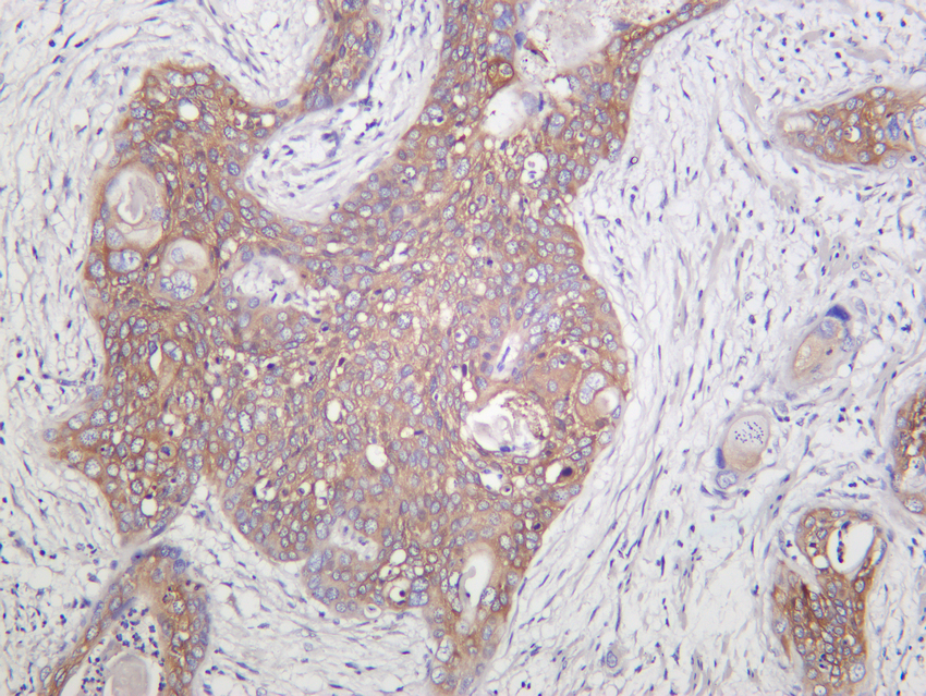
Human cervical carcinoma was stained with Anti-RIP rabbit antibody.ICC/IF
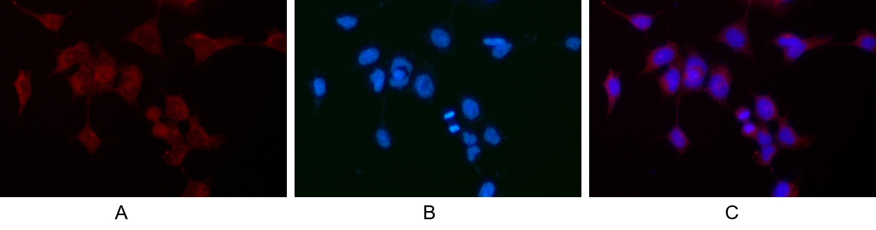
Immunofluorescence analysis of HEK293. Picture A: RIP antibody (red). Picture B: DAPI (blue). Picture C: Merge of A+B.IF-P
human cervical cancer was stained with Anti-RIP rabbit antibody.| Product Name | RIP Rabbit mAb |
|---|---|
| Antibody Type | Primary Antibodies |
| Clonality | monoclonal |
|---|---|
| Isotype | IgG |
| Host Species | Rabbit |
| Tested Applications | ICC/IFIF-PIHCWB |
| WB:1:1000-5000 IHC:1:200-1000 ICC/IF:1:200-1:1000 IF-P:1:200-1:1000 |
|
| Species Reactivity | Human |
| Concentration | 1mg/ml |
| Purification | Protein A |
| Gene Symbol | RIPK1 |
|---|---|
| Gene Synonyms | RIP RIP1 AIEFL IMD57 RIP-1 |
| Gene Full Name | receptor interacting serine/threonine kinase 1 |
| Gene Summary | This gene encodes a member of the receptor-interacting protein (RIP) family of serine/threonine protein kinases. The encoded protein plays a role in inflammation and cell death in response to tissue damage, pathogen recognition, and as part of developmental regulation. RIPK1/RIPK3 kinase-mediated necrosis is referred to as necroptosis. Genetic disruption of this gene in mice results in death shortly after birth. [provided by RefSeq, Aug 2017] |
| Molecular Weight(MW) | 76kDa |
| Cellular Localization | Cytoplasmic, Membranous. |
WB
Western blot analysis of lysates from HAP1 WT and knockout cell , (Green) primary antibody was diluted at 1:5000, 4°over night, Dylight 800 secondary antibody was diluted at 1:10000, 37° 1hour. (Red) GAPDH Monoclonal Antibody was diluted at 1:5000 as loading control, 4° over night, Dylight 680 secondary antibody was diluted at 1:10000, 37° 1hour.
WB
Various whole cell lysates were separated by 4-20% SDS-PAGE, and the membrane was blotted with anti-RIP antibody. The HRP-conjugated Goat anti-Rabbit IgG(H + L) antibody was used to detect the antibody. Lane 1: HEK293, Lane 2: Hela, Lane 3: Jurkat, Lane 4: Raji.
IHC
Rat kidney was stained with Anti-RIP rabbit antibody.
IHC
Human cervical carcinoma was stained with Anti-RIP rabbit antibody.
ICC/IF
Immunofluorescence analysis of HEK293. Picture A: RIP antibody (red). Picture B: DAPI (blue). Picture C: Merge of A+B.
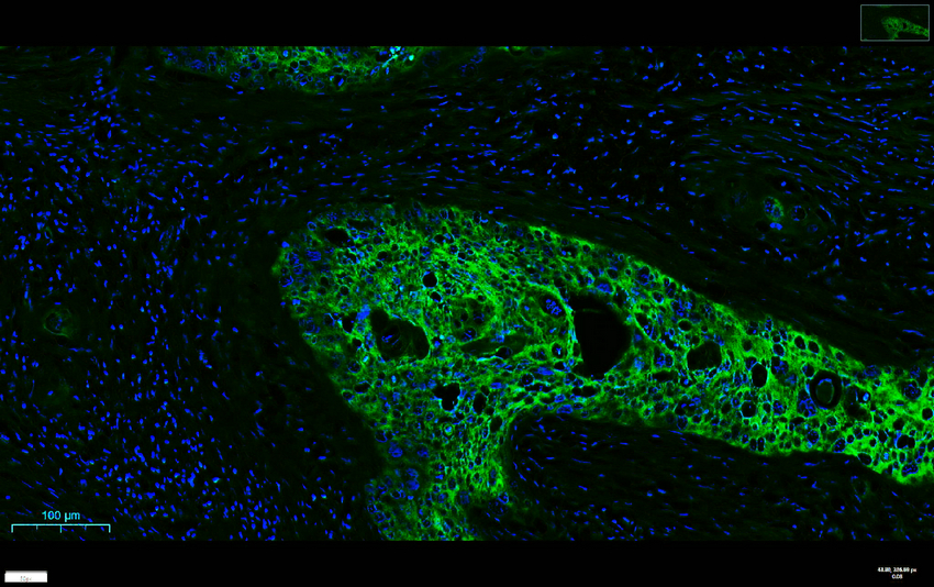
IF-P
human cervical cancer was stained with Anti-RIP rabbit antibody.| Application Notes | WB:1:1000-5000 IHC:1:200-1000 ICC/IF:1:200-1:1000 IF-P:1:200-1:1000 |
|---|
| Form | Liquid |
|---|---|
| Storage Instructions | -15°C to -25°C/1 year(Do not lower than -25°C) |
| Storage Buffer | PBS, 50% glycerol, 0.05% Proclin 300, 0.05%BSA. |


 Data sheet for OM644290