WB
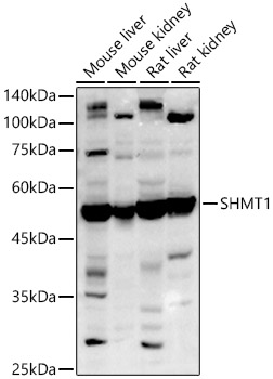
Western blot analysis of various lysates using SHMT1 Rabbit pAb at 1:1000 dilution. Secondary antibody: HRP-conjugated Goat anti-Rabbit IgG (H+L) at 1:10000 dilution. Lysates/proteins: 25μg per lane. Blocking buffer: 3% nonfat dry milk in TBST. Detection: ECL Basic Kit. Exposure time: 10s.IHC
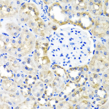
Immunohistochemistry analysis of paraffin embedded Rat kidney using SHMT1 Rabbit pAb at dilution of 1:100 (40x lens). Microwave antigen retrieval performed with 0.01M PBS Buffer (pH 7.2) prior to IHC staining.IHC
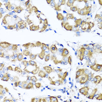
Immunohistochemistry analysis of paraffin embedded Human stomach using SHMT1 Rabbit pAb at dilution of 1:100 (40x lens). Microwave antigen retrieval performed with 0.01M PBS Buffer (pH 7.2) prior to IHC staining.ICC/IF
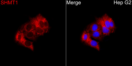
Immunofluorescence analysis of Hep G2 cells using SHMT1 Rabbit pAb at a dilution of 1:100 (40x lens). Secondary antibody:Cy3 Goat Anti-Rabbit IgG (H+L)at 1:500 dilution. Blue: DAPI for nuclear staining.IF-P
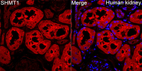
Immunofluorescence analysis of paraffin embedded Human kidney tissue using SHMT1 Rabbit pAb at a dilution of 1:100 (40x lens). Secondary antibody:Cy3 Goat Anti-Rabbit IgG (H+L)at 1:500 dilution. Blue: DAPI for nuclear staining. Perform high pressure antigen retrieval with 0.01 M citrate buffer (pH 6.0) prior to IF staining.| Product Name | SHMT1 Rabbit pAb |
|---|---|
| Antibody Type | Primary Antibodies |
| Immunogen | Recombinant fusion protein containing a sequence corresponding to amino acids 285-444 of human SHMT1 (NP_683718.1). |
| Clonality | Polyclonal |
|---|---|
| Isotype | IgG |
| Host Species | Rabbit |
| Tested Applications | ICC/IFIF-PIHCWB |
| WB:1:500-1:1000 IHC:1:50-1:200 ICC/IF:1:50-1:200 IF-P:1:50-1:200 |
|
| Species Reactivity | HumanMouseRat |
| Concentration | 1mg/ml |
| Purification | Affinity purified |
| Gene Symbol | SHMT1 |
|---|---|
| Gene Synonyms | SHMT CSHMT |
| Gene Full Name | serine hydroxymethyltransferase 1 |
| Gene Summary | This gene encodes the cytosolic form of serine hydroxymethyltransferase, a pyridoxal phosphate-containing enzyme that catalyzes the reversible conversion of serine and tetrahydrofolate to glycine and 5,10-methylene tetrahydrofolate. This reaction provides one-carbon units for synthesis of methionine, thymidylate, and purines in the cytoplasm. This gene is located within the Smith-Magenis syndrome region on chromosome 17. A pseudogene of this gene is located on the short arm of chromosome 1. Alternative splicing results in multiple transcript variants. [provided by RefSeq, Aug 2013] |
| Alternative Names | SHMT CSHMT SHMT1 |
| Molecular Weight(MW) | 50kDa |
| Cellular Localization | Cytoplasm. |
WB
Western blot analysis of various lysates using SHMT1 Rabbit pAb at 1:1000 dilution. Secondary antibody: HRP-conjugated Goat anti-Rabbit IgG (H+L) at 1:10000 dilution. Lysates/proteins: 25μg per lane. Blocking buffer: 3% nonfat dry milk in TBST. Detection: ECL Basic Kit. Exposure time: 10s.
IHC
Immunohistochemistry analysis of paraffin embedded Rat kidney using SHMT1 Rabbit pAb at dilution of 1:100 (40x lens). Microwave antigen retrieval performed with 0.01M PBS Buffer (pH 7.2) prior to IHC staining.
IHC
Immunohistochemistry analysis of paraffin embedded Human stomach using SHMT1 Rabbit pAb at dilution of 1:100 (40x lens). Microwave antigen retrieval performed with 0.01M PBS Buffer (pH 7.2) prior to IHC staining.
ICC/IF
Immunofluorescence analysis of Hep G2 cells using SHMT1 Rabbit pAb at a dilution of 1:100 (40x lens). Secondary antibody:Cy3 Goat Anti-Rabbit IgG (H+L)at 1:500 dilution. Blue: DAPI for nuclear staining.
IF-P
Immunofluorescence analysis of paraffin embedded Human kidney tissue using SHMT1 Rabbit pAb at a dilution of 1:100 (40x lens). Secondary antibody:Cy3 Goat Anti-Rabbit IgG (H+L)at 1:500 dilution. Blue: DAPI for nuclear staining. Perform high pressure antigen retrieval with 0.01 M citrate buffer (pH 6.0) prior to IF staining.| Application Notes | WB:1:500-1:1000 IHC:1:50-1:200 ICC/IF:1:50-1:200 IF-P:1:50-1:200 |
|---|
| Form | Liquid |
|---|---|
| Storage Instructions | Store at -20℃. Avoid freeze / thaw cycles. |
| Storage Buffer | PBS with 0.05% proclin300, 50% glycerol, pH7.3. |


