WB
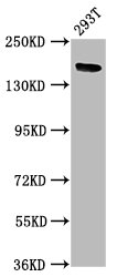
Western Blot Positive WB detected in: 293T whole cell lysate, All lanes: ITSN1 antibody at 5µg/ml, Secondary Goat polyclonal to rabbit IgG at 1/50000 dilution.IHC
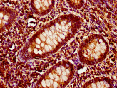
IHC image of ITSN1 Antibody diluted at 1:400 and staining in paraffin-embedded human appendix tissue performed on a Leica BondTM system. After dewaxing and hydration, antigen retrieval was mediated by high pressure in a citrate buffer (pH 6.0). Section was blocked with 10% normal goat serum 30min at RT. Then primary antibody (1% BSA) was incubated at 4°C overnight. The primary is detected by a biotinylated secondary antibody and visualized using an HRP conjugated SP system.| Product Name | Rabbit anti-ITSN1 Polyclonal antibody |
|---|---|
| Antibody Type | Primary Antibodies |
| Immunogen | Recombinant Human Intersectin-1 protein (773-916AA). |
| Clonality | Polyclonal |
|---|---|
| Isotype | IgG |
| Host Species | Rabbit |
| Tested Applications | IHCWB |
| WB:1:500-1:5000 IHC:1:200-1:500 |
|
| Species Reactivity | Human |
| Concentration | 1mg/ml |
| Purification | Protein G |
| Gene Symbol | ITSN1 |
|---|---|
| Gene Synonyms | ITSN SH3D1A SH3P17 |
| Gene Full Name | intersectin 1 |
| Gene Summary | The protein encoded by this gene is a cytoplasmic membrane-associated protein that indirectly coordinates endocytic membrane traffic with the actin assembly machinery. In addition, the encoded protein may regulate the formation of clathrin-coated vesicles and could be involved in synaptic vesicle recycling. This protein has been shown to interact with dynamin, CDC42, SNAP23, SNAP25, SPIN90, EPS15, EPN1, EPN2, and STN2. Multiple transcript variants encoding different isoforms have been found for this gene, but the full-length nature of only two of them have been characterized so far. [provided by RefSeq, Jul 2008] |
| Alternative Names | Human intersectin SH3 domain containing protein SH3P17 antibody Intersectin 1 SH3 domain protein antibody intersectin 1 short form variant 3 antibody intersectin 1 short form variant 11 antibody Intersectin short form antibody intersectin short variant 12 antibody Intersectin-1 antibody Intersectin1 antibody ITSN 1 antibody ITSN antibody itsn1 antibody ITSN1_HUMAN antibody MGC134948 antibody MGC134949 antibody OTTHUMP00000067855 antibody OTTHUMP00000067856 antibody OTTHUMP00000067857 antibody OTTHUMP00000194810 antibody OTTHUMP00000194811 antibody OTTHUMP00000194812 antibody OTTHUMP00000206688 antibody SH3 domain containing protein 1A antibody SH3 domain protein 1A antibody SH3 domain-containing protein 1A antibody SH3D1A antibody SH3P17 antibody Src homology 3 domain containing protein antibody |
| Molecular Weight(MW) | 196kDa |
| Cellular Localization | Cell membrane,Cell projection,Coated pit,Cytoplasm,Cytoplasmic vesicle,Endosome,Membrane,Nucleus,Synapse,Synaptosome. |
WB
Western Blot Positive WB detected in: 293T whole cell lysate, All lanes: ITSN1 antibody at 5µg/ml, Secondary Goat polyclonal to rabbit IgG at 1/50000 dilution.
IHC
IHC image of ITSN1 Antibody diluted at 1:400 and staining in paraffin-embedded human appendix tissue performed on a Leica BondTM system. After dewaxing and hydration, antigen retrieval was mediated by high pressure in a citrate buffer (pH 6.0). Section was blocked with 10% normal goat serum 30min at RT. Then primary antibody (1% BSA) was incubated at 4°C overnight. The primary is detected by a biotinylated secondary antibody and visualized using an HRP conjugated SP system.| Application Notes | WB:1:500-1:5000 IHC:1:200-1:500 |
|---|
| Form | Liquid |
|---|---|
| Storage Instructions | Upon receipt, store at -20°C or -80°C. Avoid repeated freeze. |
| Storage Buffer | Preservative: 0.03% Proclin 300,Constituents: 50% Glycerol, 0.01M PBS, pH 7.4 |


