WB
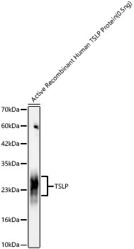
Western blot analysis of Active Recombinant Human TSLP Protein using TSLP Rabbit mAb at 1:1000 dilution. Secondary antibody: HRP-conjugated Goat anti-Rabbit IgG (H+L)at 1:10000 dilution. Lysates/proteins: 0.5ng per lane. Blocking buffer: 3% nonfat dry milk in TBST. Detection: ECL Basic Kit. Exposure time: 45s.IHC
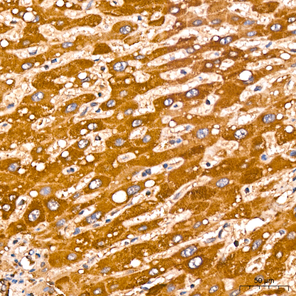
Immunohistochemistry analysis of paraffin embedded Human liver tissue using TSLP Rabbit mAb at a dilution of 1:200 (40x lens). High pressure antigen retrieval performed with 0.01M Citrate Bufferr (pH 6.0) prior to IHC staining.ICC/IF
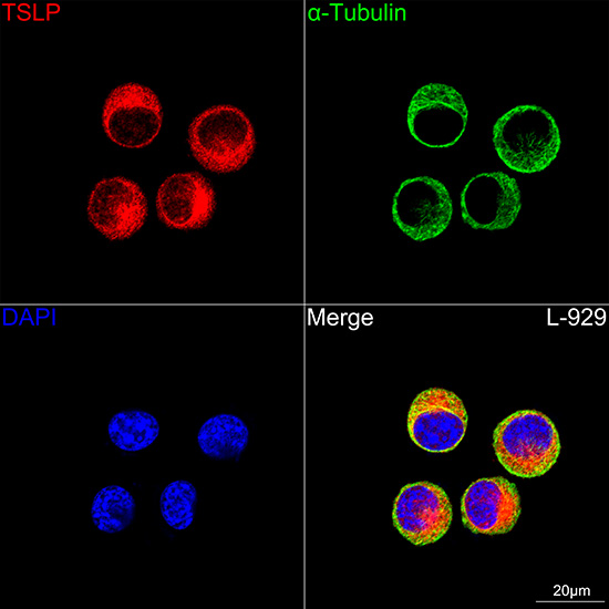
Confocal imaging of L-929 cells using TSLP Rabbit mAb (dilution 1:200) followed by a further incubation with Cy3 Goat Anti-Rabbit IgG (H+L) (dilution 1:500) (Red). The cells were counterstained with α-Tubulin Mouse mAb (dilution 1:400) followed by incubation with Omnimabs® 488 Goat Anti-Mouse IgG(H&L) Ab (dilution 1:500) (Green). DAPI was used for nuclear staining (Blue). Objective: 100x.ICC/IF
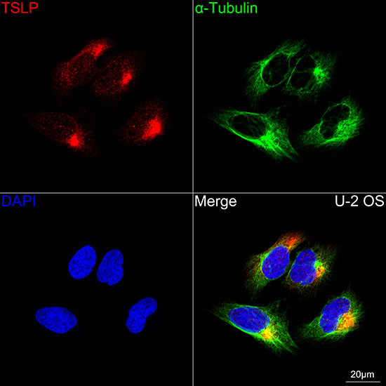
Confocal imaging of U-2 OS cells using TSLP Rabbit mAb (dilution 1:200) followed by a further incubation with Cy3 Goat Anti-Rabbit IgG (H+L) (dilution 1:500) (Red). The cells were counterstained with α-Tubulin Mouse mAb (dilution 1:400) followed by incubation with Omnimabs® 488 Goat Anti-Mouse IgG(H&L) Ab (dilution 1:500) (Green). DAPI was used for nuclear staining (Blue). Objective: 100x.IF-P
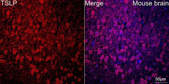
Confocal imaging of paraffin-embedded Mouse brain tissue using TSLP Rabbit mAb (dilution 1:200) followed by a further incubation with Cy3 Goat Anti-Rabbit IgG (H+L) (dilution 1:500) (Red). DAPI was used for nuclear staining (Blue). Objective: 40x.| Product Name | TSLP Rabbit mAb |
|---|---|
| Antibody Type | Primary Antibodies |
| Immunogen | Recombinant fusion protein containing a sequence corresponding to amino acids 29-159 of human TSLP(NP_149024.1). |
| Clonality | Monoclonal |
|---|---|
| Isotype | IgG |
| Host Species | Rabbit |
| Tested Applications | ICC/IFIF-PIHCWB |
| WB:1:500-1:1000 IHC:1:50-1:200 ICC/IF:1:50-1:200 IF-P:1:50-1:200 |
|
| Species Reactivity | HumanMouse |
| Concentration | 1mg/ml |
| Purification | Affinity purified |
| Gene Symbol | TSLP |
|---|---|
| Gene Full Name | thymic stromal lymphopoietin |
| Gene Summary | This gene encodes a hemopoietic cytokine proposed to signal through a heterodimeric receptor complex composed of the thymic stromal lymphopoietin receptor and the IL-7R alpha chain. It mainly impacts myeloid cells and induces the release of T cell-attracting chemokines from monocytes and enhances the maturation of CD11c(+) dendritic cells. The protein promotes T helper type 2 (TH2) cell responses that are associated with immunity in various inflammatory diseases, including asthma, allergic inflammation and chronic obstructive pulmonary disease. The protein is therefore considered a potential therapeutic target for the treatment of such diseases. In addition, the shorter (predominant) isoform is an antimicrobial protein, displaying antibacterial and antifungal activity against B. cereus, E. coli, E. faecalis, S. mitis, S. epidermidis, and C. albicans. Alternative splicing of this gene results in multiple transcript variants. [provided by RefSeq, Jul 2020] |
| Alternative Names | TSLP |
| Molecular Weight(MW) | 19kDa(Observed MW: 20-30kDa Recombinant Protein) |
| Cellular Localization | Secreted. |
WB
Western blot analysis of Active Recombinant Human TSLP Protein using TSLP Rabbit mAb at 1:1000 dilution. Secondary antibody: HRP-conjugated Goat anti-Rabbit IgG (H+L)at 1:10000 dilution. Lysates/proteins: 0.5ng per lane. Blocking buffer: 3% nonfat dry milk in TBST. Detection: ECL Basic Kit. Exposure time: 45s.
IHC
Immunohistochemistry analysis of paraffin embedded Human liver tissue using TSLP Rabbit mAb at a dilution of 1:200 (40x lens). High pressure antigen retrieval performed with 0.01M Citrate Bufferr (pH 6.0) prior to IHC staining.
ICC/IF
Confocal imaging of L-929 cells using TSLP Rabbit mAb (dilution 1:200) followed by a further incubation with Cy3 Goat Anti-Rabbit IgG (H+L) (dilution 1:500) (Red). The cells were counterstained with α-Tubulin Mouse mAb (dilution 1:400) followed by incubation with Omnimabs® 488 Goat Anti-Mouse IgG(H&L) Ab (dilution 1:500) (Green). DAPI was used for nuclear staining (Blue). Objective: 100x.
ICC/IF
Confocal imaging of U-2 OS cells using TSLP Rabbit mAb (dilution 1:200) followed by a further incubation with Cy3 Goat Anti-Rabbit IgG (H+L) (dilution 1:500) (Red). The cells were counterstained with α-Tubulin Mouse mAb (dilution 1:400) followed by incubation with Omnimabs® 488 Goat Anti-Mouse IgG(H&L) Ab (dilution 1:500) (Green). DAPI was used for nuclear staining (Blue). Objective: 100x.
IF-P
Confocal imaging of paraffin-embedded Mouse brain tissue using TSLP Rabbit mAb (dilution 1:200) followed by a further incubation with Cy3 Goat Anti-Rabbit IgG (H+L) (dilution 1:500) (Red). DAPI was used for nuclear staining (Blue). Objective: 40x.| Application Notes | WB:1:500-1:1000 IHC:1:50-1:200 ICC/IF:1:50-1:200 IF-P:1:50-1:200 |
|---|
| Form | Liquid |
|---|---|
| Storage Instructions | Store at -20℃. Avoid freeze / thaw cycles. |
| Storage Buffer | PBS with 0.05% proclin300, 50% glycerol, pH7.3. |


