WB
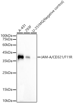
Western blot analysis of various lysates, using JAM-A/CD321/F11R Rabbit mAb at 1:8000 dilution. Secondary antibody: HRP-conjugated Goat anti-Rabbit IgG (H+L) at 1:10000 dilution. Lysates/proteins: 25μg per lane. Blocking buffer: 3% nonfat dry milk in TBST. Detection: ECL Basic Kit. Exposure time: 90s.ICC/IF
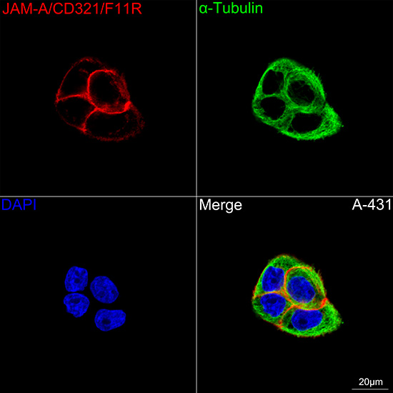
Confocal imaging of A-431 cells using JAM A/CD321/F11R Rabbit mAb (dilution 1:200) followed by a further incubation with Cy3 Goat Anti-Rabbit IgG (H+L) (dilution 1:500) (Red). The cells were counterstained with α-Tubulin Mouse mAb (dilution 1:400) followed by incubation with Omnimabs® 488 Goat Anti-Mouse IgG(H&L) Ab (dilution 1:500) (Green). DAPI was used for nuclear staining (Blue). Objective: 100x.| Product Name | JAM-A/CD321/F11R Rabbit mAb |
|---|---|
| Antibody Type | Primary Antibodies |
| Immunogen | Recombinant protein. |
| Clonality | Monoclonal |
|---|---|
| Isotype | IgG |
| Host Species | Rabbit |
| Tested Applications | ICC/IFWB |
| WB:1:4000-1:8000 ICC/IF:1:200-1:800 |
|
| Species Reactivity | Human |
| Concentration | 1mg/ml |
| Purification | Affinity purified |
| Gene Symbol | F11R |
|---|---|
| Gene Synonyms | JAM KAT JAM1 JAMA JCAM CD321 PAM-1 |
| Gene Full Name | F11 receptor |
| Gene Summary | Tight junctions represent one mode of cell-to-cell adhesion in epithelial or endothelial cell sheets, forming continuous seals around cells and serving as a physical barrier to prevent solutes and water from passing freely through the paracellular space. The protein encoded by this immunoglobulin superfamily gene member is an important regulator of tight junction assembly in epithelia. In addition, the encoded protein can act as (1) a receptor for reovirus, (2) a ligand for the integrin LFA1, involved in leukocyte transmigration, and (3) a platelet receptor. Multiple 5' alternatively spliced variants, encoding the same protein, have been identified but their biological validity has not been established. [provided by RefSeq, Jul 2008] |
| Alternative Names | JAM KAT JAM1 JAMA JCAM CD321 PAM-1 JAM-A/CD321/F11R |
| Molecular Weight(MW) | 33kDa(Observed MW 38kDa) |
| Cellular Localization | Cell junction, Cell membrane, Single-pass type I membrane protein, tight junction. |
WB
Western blot analysis of various lysates, using JAM-A/CD321/F11R Rabbit mAb at 1:8000 dilution. Secondary antibody: HRP-conjugated Goat anti-Rabbit IgG (H+L) at 1:10000 dilution. Lysates/proteins: 25μg per lane. Blocking buffer: 3% nonfat dry milk in TBST. Detection: ECL Basic Kit. Exposure time: 90s.
ICC/IF
Confocal imaging of A-431 cells using JAM A/CD321/F11R Rabbit mAb (dilution 1:200) followed by a further incubation with Cy3 Goat Anti-Rabbit IgG (H+L) (dilution 1:500) (Red). The cells were counterstained with α-Tubulin Mouse mAb (dilution 1:400) followed by incubation with Omnimabs® 488 Goat Anti-Mouse IgG(H&L) Ab (dilution 1:500) (Green). DAPI was used for nuclear staining (Blue). Objective: 100x.| Application Notes | WB:1:4000-1:8000 ICC/IF:1:200-1:800 |
|---|
| Form | Liquid |
|---|---|
| Storage Instructions | Store at -20℃. Avoid freeze / thaw cycles. |
| Storage Buffer | PBS with 0.09% Sodium azide, 0.05% BSA, 50% glycerol, pH7.3. |


