WB
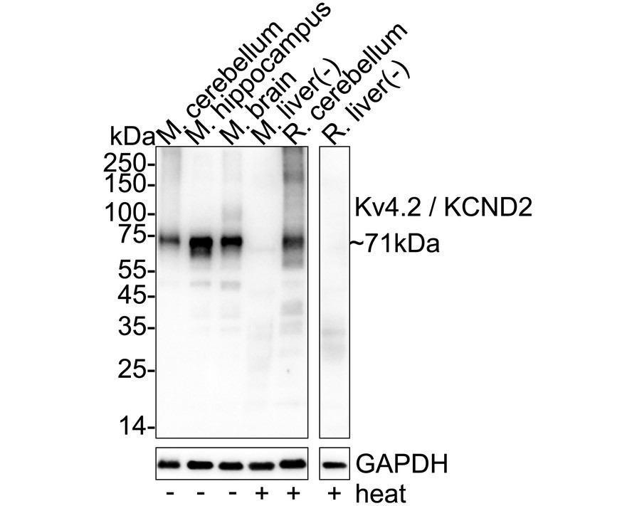
Western blot analysis of Kv4.2 / KCND2 on different lysates with Mouse anti-Kv4.2 / KCND2 antibody at 1/5,000 dilution. Lane 1: Mouse cerebellum tissue lysate (no heat), Lane 2: Mouse hippocampus tissue lysate (no heat), Lane 3: Mouse brain tissue lysate (no heat), Lane 4: Mouse liver tissue lysate (negative), Lane 5: Rat cerebellum tissue lysate, Lane 6: Rat liver tissue lysate (negative), Notice: no heat means the lysate is not boiled. Lysates/proteins at 40 µg/Lane. Exposure time: 1 minute 2 seconds; 4-20% SDS-PAGE gel. Proteins were transferred to a PVDF membrane and blocked with 5% NFDM/TBST for 1 hour at room temperature. The primary antibody at 1/5,000 dilution was used in primary antibody dilution at 4℃ overnight. Goat Anti-Mouse IgG - HRP Secondary Antibody at 1/50,000 dilution was used for 1 hour at room temperature.IHC
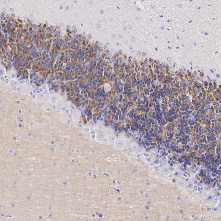
Immunohistochemical analysis of paraffin-embedded mouse cerebellum tissue with Mouse anti-Kv4.2 / KCND2 antibody at 1/1,000 dilution. The section was pre-treated using heat mediated antigen retrieval with Tris-EDTA buffer (pH 9.0) for 20 minutes. The tissues were blocked in 1% BSA for 20 minutes at room temperature, washed with ddH2O and PBS, and then probed with the primary antibody at 1/1,000 dilution for 1 hour at room temperature. The detection was performed using an HRP conjugated compact polymer system. DAB was used as the chromogen. Tissues were counterstained with hematoxylin and mounted with DPX.IHC
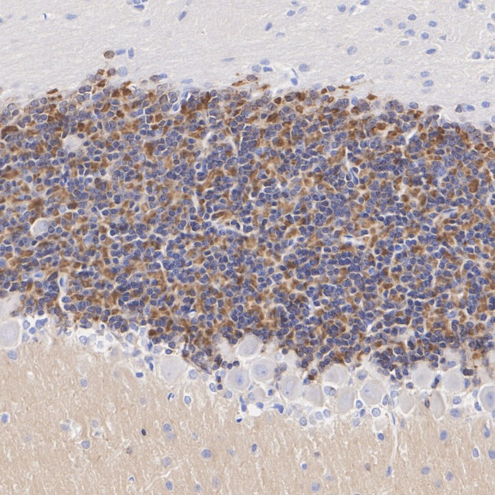
Immunohistochemical analysis of paraffin-embedded rat cerebellum tissue with Mouse anti-Kv4.2 / KCND2 antibody at 1/1,000 dilution. The section was pre-treated using heat mediated antigen retrieval with Tris-EDTA buffer (pH 9.0) for 20 minutes. The tissues were blocked in 1% BSA for 20 minutes at room temperature, washed with ddH2O and PBS, and then probed with the primary antibody at 1/1,000 dilution for 1 hour at room temperature. The detection was performed using an HRP conjugated compact polymer system. DAB was used as the chromogen. Tissues were counterstained with hematoxylin and mounted with DPX.IF-F
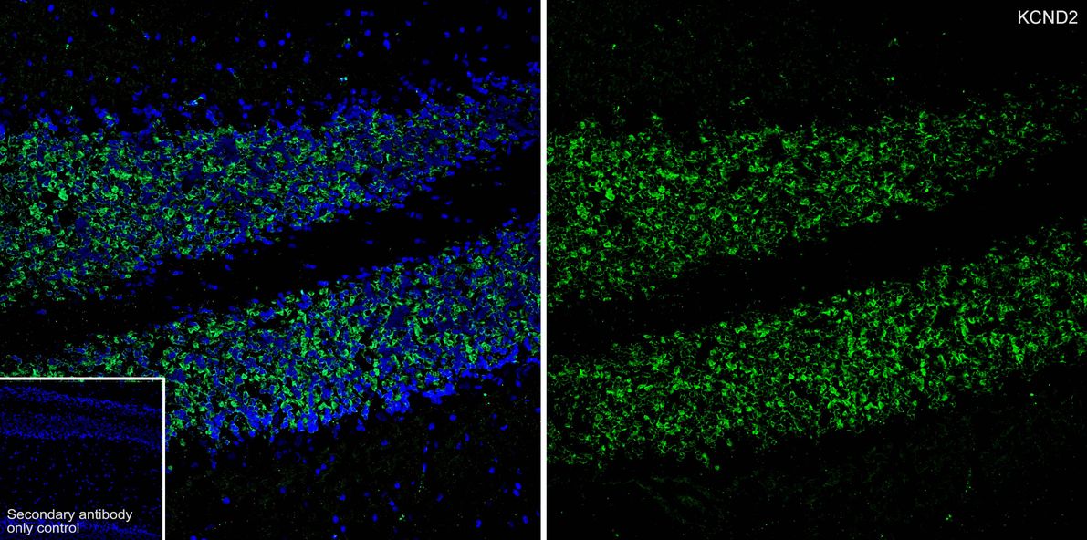
Species: Rat, Site: cerebellum, Sample: Frozen section, Antibody concentration: 1/500, Antigen retrieval: Not required.| Product Name | Kv4.2 / KCND2 Recombinant Mouse Monoclonal Antibody |
|---|---|
| Antibody Type | Primary Antibodies |
| Clonality | monoclonal |
|---|---|
| Isotype | IgG1 |
| Host Species | Mouse |
| Tested Applications | IF-FIHCWB |
| WB:1:5000 IHC:1:1000 IF-F:1:500 |
|
| Species Reactivity | MouseRat |
| Concentration | 1mg/ml |
| Purification | Protein A |
| Gene Symbol | Kcnd2 |
|---|---|
| Gene Synonyms | Kv4.2 Gm52855 |
| Gene Full Name | potassium voltage-gated channel, Shal-related family, member 2 |
| Gene Summary | Enables A-type (transient outward) potassium channel activity and voltage-gated monoatomic ion channel activity involved in regulation of postsynaptic membrane potential. Involved in several processes, including locomotor rhythm; neuronal action potential; and potassium ion transmembrane transport. Located in dendritic spine; neuronal cell body; and plasma membrane. Part of voltage-gated potassium channel complex. Is active in GABA-ergic synapse; glutamatergic synapse; and postsynaptic specialization membrane. Is expressed in several structures, including heart; hindlimb digit; metanephros; nervous system; and sensory organ. Orthologous to human KCND2 (potassium voltage-gated channel subfamily D member 2). [provided by Alliance of Genome Resources, Jul 2025] |
| Alternative Names | KCD2 antibody KCND 2 antibody KCND2 antibody KCND2_HUMAN antibody KIAA1044 antibody MGC119702 antibody MGC119703 antibody Potassium voltage gated channel Shal related subfamily member 2 antibody Potassium voltage-gated channel subfamily D member 2 antibody RK 5 antibody KCD2 antibody KCND 2 antibody KCND2 antibody KCND2_HUMAN antibody KIAA1044 antibody MGC119702 antibody MGC119703 antibody Potassium voltage gated channel Shal related subfamily member 2 antibody Potassium voltage-gated channel subfamily D member 2 antibody RK 5 antibody RK5 antibody Voltage gated potassium channel Kv4.2 antibody Voltage gated potassium channel subunit Kv4.2 antibody Voltage sensitive potassium channel antibody Voltage-gated potassium channel subunit Kv4.2 antibody |
| Molecular Weight(MW) | 71kDa |
| Cellular Localization | Cell membrane, Cell projection, dendrite, Synapse, Perikaryon, Postsynaptic cell membrane, Cell projection, Cell junction. |
WB
Western blot analysis of Kv4.2 / KCND2 on different lysates with Mouse anti-Kv4.2 / KCND2 antibody at 1/5,000 dilution. Lane 1: Mouse cerebellum tissue lysate (no heat), Lane 2: Mouse hippocampus tissue lysate (no heat), Lane 3: Mouse brain tissue lysate (no heat), Lane 4: Mouse liver tissue lysate (negative), Lane 5: Rat cerebellum tissue lysate, Lane 6: Rat liver tissue lysate (negative), Notice: no heat means the lysate is not boiled. Lysates/proteins at 40 µg/Lane. Exposure time: 1 minute 2 seconds; 4-20% SDS-PAGE gel. Proteins were transferred to a PVDF membrane and blocked with 5% NFDM/TBST for 1 hour at room temperature. The primary antibody at 1/5,000 dilution was used in primary antibody dilution at 4℃ overnight. Goat Anti-Mouse IgG - HRP Secondary Antibody at 1/50,000 dilution was used for 1 hour at room temperature.
IHC
Immunohistochemical analysis of paraffin-embedded mouse cerebellum tissue with Mouse anti-Kv4.2 / KCND2 antibody at 1/1,000 dilution. The section was pre-treated using heat mediated antigen retrieval with Tris-EDTA buffer (pH 9.0) for 20 minutes. The tissues were blocked in 1% BSA for 20 minutes at room temperature, washed with ddH2O and PBS, and then probed with the primary antibody at 1/1,000 dilution for 1 hour at room temperature. The detection was performed using an HRP conjugated compact polymer system. DAB was used as the chromogen. Tissues were counterstained with hematoxylin and mounted with DPX.
IHC
Immunohistochemical analysis of paraffin-embedded rat cerebellum tissue with Mouse anti-Kv4.2 / KCND2 antibody at 1/1,000 dilution. The section was pre-treated using heat mediated antigen retrieval with Tris-EDTA buffer (pH 9.0) for 20 minutes. The tissues were blocked in 1% BSA for 20 minutes at room temperature, washed with ddH2O and PBS, and then probed with the primary antibody at 1/1,000 dilution for 1 hour at room temperature. The detection was performed using an HRP conjugated compact polymer system. DAB was used as the chromogen. Tissues were counterstained with hematoxylin and mounted with DPX.
IF-F
Species: Rat, Site: cerebellum, Sample: Frozen section, Antibody concentration: 1/500, Antigen retrieval: Not required.| Application Notes | WB:1:5000 IHC:1:1000 IF-F:1:500 |
|---|
| Form | Liquid |
|---|---|
| Storage Instructions | Store at +4℃ after thawing. Aliquot store at -20℃. Avoid repeated freeze / thaw cycles. |
| Storage Buffer | PBS (pH7.4), 0.1% BSA, 40% Glycerol. Preservative: 0.05% Sodium Azide. |


