WB
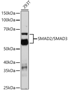
Western blot analysis of various lysates using SMAD2/SMAD3 Rabbit pAb at 1:1000 dilution. Secondary antibody: HRP-conjugated Goat anti-Rabbit IgG (H+L) at 1:10000 dilution. Lysates/proteins: 25μg per lane. Blocking buffer: 3% nonfat dry milk in TBST. Detection: ECL Enhanced Kit. Exposure time: 3min.IHC
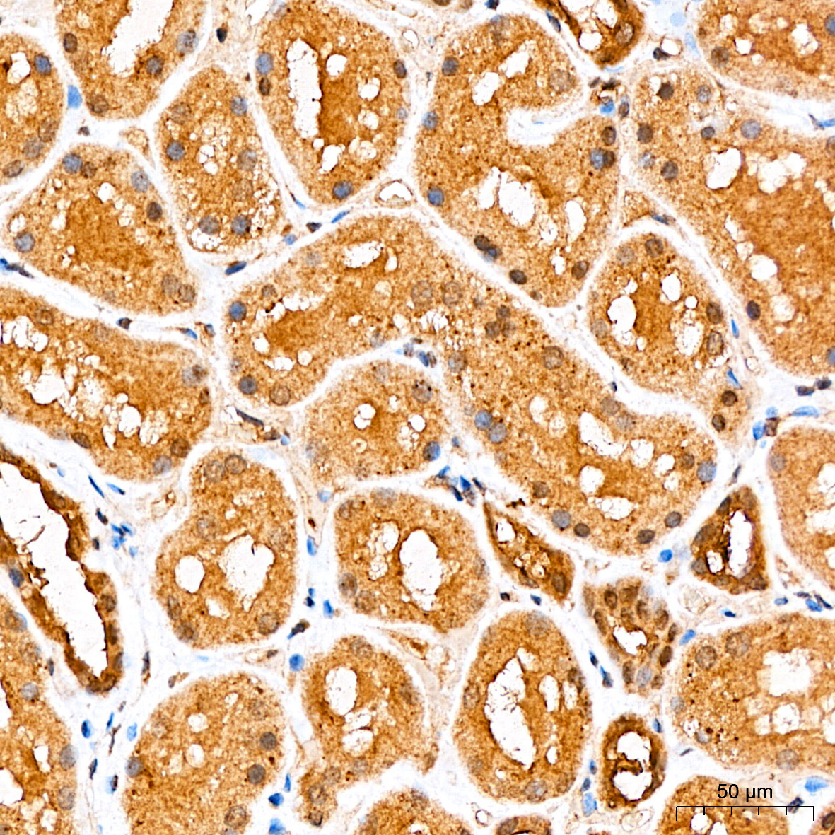
Immunohistochemistry analysis of paraffin embedded Human kidney tissue using SMAD2/SMAD3 Rabbit pAb at a dilution of 1:100 (40x lens). High pressure antigen retrieval performed with 0.01M Citrate Buffer(pH 6.0) prior to IHC staining.IHC
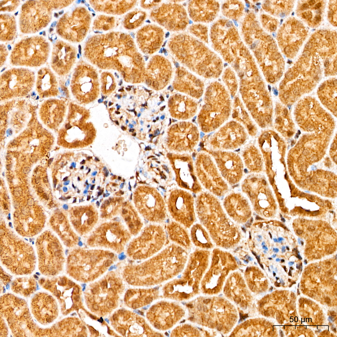
Immunohistochemistry analysis of paraffin embedded Mouse kidney tissue using SMAD2/SMAD3 Rabbit pAb at a dilution of 1:100 (40x lens). High pressure antigen retrieval performed with 0.01M Citrate Buffer(pH 6.0) prior to IHC staining.IHC
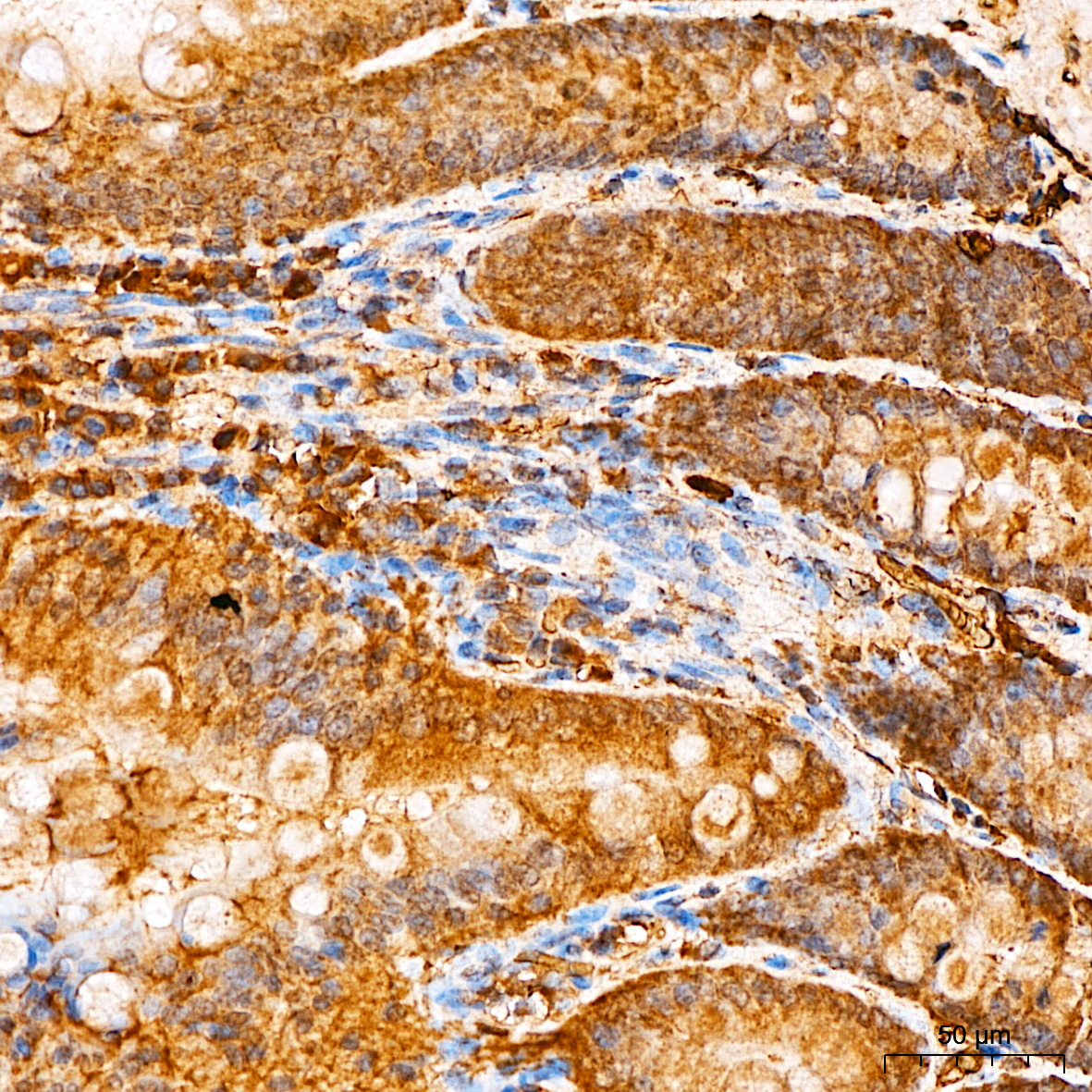
Immunohistochemistry analysis of paraffin embedded Rat intestine tissue using SMAD2/SMAD3 Rabbit pAb at a dilution of 1:100 (40x lens). High pressure antigen retrieval performed with 0.01M Citrate Buffer(pH 6.0) prior to IHC staining.ICC/IF
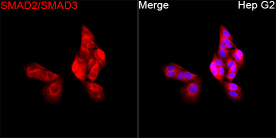
Immunofluorescence analysis of Hep G2 cells using SMAD2/SMAD3 Rabbit pAb at a dilution of 1:100 (40x lens). Secondary antibody: Cy3-conjugated Goat anti-Rabbit IgG (H+L) at 1:500 dilution. Blue: DAPI for nuclear staining.| Product Name | SMAD2/SMAD3 Rabbit pAb |
|---|---|
| Antibody Type | Primary Antibodies |
| Immunogen | Synthetic peptide. |
| Clonality | Polyclonal |
|---|---|
| Isotype | IgG |
| Host Species | Rabbit |
| Tested Applications | ICC/IFIHCWB |
| WB:1:500-1:2000 IHC:1:50-1:200 ICC/IF:1:50-1:200 |
|
| Species Reactivity | HumanMouseRat |
| Concentration | 1mg/ml |
| Purification | Affinity purified |
| Gene Symbol | SMAD2/SMAD3 |
|---|---|
| Gene Synonyms | JV18 LDS6 CHTD8 MADH2 MADR2 JV18-1 hMAD-2 hSMAD2 LDS3 mad3 LDS1C MADH3 JV15-2 hMAD-3 hSMAD3 HSPC193 HsT17436 |
| Gene Full Name | SMAD family member 2/3 |
| Gene Summary | The protein encoded by this gene belongs to the SMAD, a family of proteins similar to the gene products of the Drosophila gene 'mothers against decapentaplegic' (Mad) and the C. elegans gene Sma. SMAD proteins are signal transducers and transcriptional modulators that mediate multiple signaling pathways. This protein mediates the signal of the transforming growth factor (TGF)-beta, and thus regulates multiple cellular processes, such as cell proliferation, apoptosis, and differentiation. This protein is recruited to the TGF-beta receptors through its interaction with the SMAD anchor for receptor activation (SARA) protein. In response to TGF-beta signal, this protein is phosphorylated by the TGF-beta receptors. The phosphorylation induces the dissociation of this protein with SARA and the association with the family member SMAD4. The association with SMAD4 is important for the translocation of this protein into the nucleus, where it binds to target promoters and forms a transcription repressor complex with other cofactors. This protein can also be phosphorylated by activin type 1 receptor kinase, and mediates the signal from the activin. Alternatively spliced transcript variants have been observed for this gene. [provided by RefSeq, May 2012] |
| Alternative Names | HSPC193 HsT17436 JV15-2 LDS1C LDS3 MADH3 SMAD2/SMAD3 |
| Molecular Weight(MW) | 52kDa/60kDa |
| Cellular Localization | Cytoplasm, Nucleus. |
WB
Western blot analysis of various lysates using SMAD2/SMAD3 Rabbit pAb at 1:1000 dilution. Secondary antibody: HRP-conjugated Goat anti-Rabbit IgG (H+L) at 1:10000 dilution. Lysates/proteins: 25μg per lane. Blocking buffer: 3% nonfat dry milk in TBST. Detection: ECL Enhanced Kit. Exposure time: 3min.
IHC
Immunohistochemistry analysis of paraffin embedded Human kidney tissue using SMAD2/SMAD3 Rabbit pAb at a dilution of 1:100 (40x lens). High pressure antigen retrieval performed with 0.01M Citrate Buffer(pH 6.0) prior to IHC staining.
IHC
Immunohistochemistry analysis of paraffin embedded Mouse kidney tissue using SMAD2/SMAD3 Rabbit pAb at a dilution of 1:100 (40x lens). High pressure antigen retrieval performed with 0.01M Citrate Buffer(pH 6.0) prior to IHC staining.
IHC
Immunohistochemistry analysis of paraffin embedded Rat intestine tissue using SMAD2/SMAD3 Rabbit pAb at a dilution of 1:100 (40x lens). High pressure antigen retrieval performed with 0.01M Citrate Buffer(pH 6.0) prior to IHC staining.
ICC/IF
Immunofluorescence analysis of Hep G2 cells using SMAD2/SMAD3 Rabbit pAb at a dilution of 1:100 (40x lens). Secondary antibody: Cy3-conjugated Goat anti-Rabbit IgG (H+L) at 1:500 dilution. Blue: DAPI for nuclear staining.| Application Notes | WB:1:500-1:2000 IHC:1:50-1:200 ICC/IF:1:50-1:200 |
|---|
| Form | Liquid |
|---|---|
| Storage Instructions | Store at -20℃. Avoid freeze / thaw cycles. |
| Storage Buffer | PBS containing 50% glycerol and 0.05% BSA, preserved with proclin300 or sodium azide (as specified on the Certificate of Analysis), pH 7.3. |


