WB
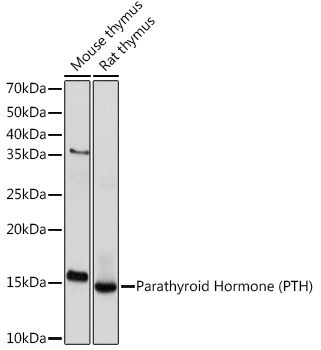
Western blot analysis of various lysates using Parathyroid Hormone (PTH) Rabbit mAb at 1:1000 dilution. Secondary antibody: HRP-conjugated Goat anti-Rabbit IgG (H+L) at 1:10000 dilution. Lysates/proteins: 25μg per lane. Blocking buffer: 3% nonfat dry milk in TBST. Detection: ECL Basic Kit. Exposure time: 10s.IF-P
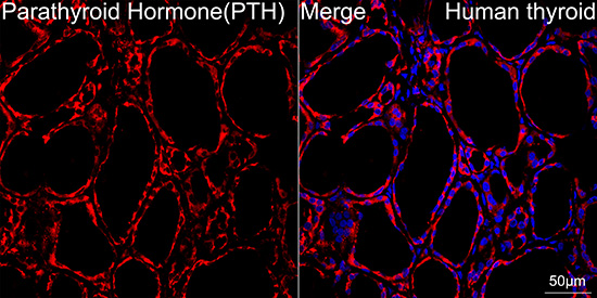
Confocal imaging of paraffin-embedded Human thyroid tissue using Parathyroid Hormone (PTH) Rabbit mAb (dilution 1:200) followed by a further incubation with Cy3 Goat Anti-Rabbit IgG (H+L) (dilution 1:500) (Red). DAPI was used for nuclear staining (Blue). Objective: 40x. Perform high pressure antigen retrieval with 0.01 M citrate buffer (pH 6.0) prior to IF staining.IF-P
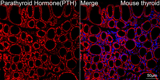
Confocal imaging of paraffin-embedded Mouse thyroid tissue using Parathyroid Hormone (PTH) Rabbit mAb (dilution 1:200) followed by a further incubation with Cy3 Goat Anti-Rabbit IgG (H+L) (dilution 1:500) (Red). DAPI was used for nuclear staining (Blue). Objective: 40x. Perform high pressure antigen retrieval with 0.01 M citrate buffer (pH 6.0) prior to IF staining.| Product Name | Parathyroid Hormone (PTH) Rabbit mAb |
|---|---|
| Antibody Type | Primary Antibodies |
| Immunogen | Synthetic peptide. |
| Clonality | Monoclonal |
|---|---|
| Isotype | IgG |
| Host Species | Rabbit |
| Tested Applications | IF-PWB |
| WB:1:1000-1:3000 IF-P:1:200-1:2000 |
|
| Species Reactivity | HumanMouseRat |
| Concentration | 1mg/ml |
| Purification | Affinity purified |
| Gene Symbol | PTH |
|---|---|
| Gene Synonyms | FIH1 PTH1 |
| Gene Full Name | parathyroid hormone |
| Gene Summary | This gene encodes a member of the parathyroid family of proteins. The encoded preproprotein is proteolytically processed to generate a protein that binds to the parathyroid hormone/parathyroid hormone-related peptide receptor and regulates blood calcium and phosphate levels. Excess production of the encoded protein, known as hyperparathyroidism, can result in hypercalcemia and kidney stones. On the other hand, defective processing of the encoded protein may lead to hypoparathyroidism, which can result in hypocalcemia and numbness. Alternative splicing results in multiple transcript variants. [provided by RefSeq, Oct 2015] |
| Alternative Names | FIH1 PTH1 Parathyroid Hormone (PTH) |
| Molecular Weight(MW) | 13kDa |
| Cellular Localization | Secreted. |
WB
Western blot analysis of various lysates using Parathyroid Hormone (PTH) Rabbit mAb at 1:1000 dilution. Secondary antibody: HRP-conjugated Goat anti-Rabbit IgG (H+L) at 1:10000 dilution. Lysates/proteins: 25μg per lane. Blocking buffer: 3% nonfat dry milk in TBST. Detection: ECL Basic Kit. Exposure time: 10s.
IF-P
Confocal imaging of paraffin-embedded Human thyroid tissue using Parathyroid Hormone (PTH) Rabbit mAb (dilution 1:200) followed by a further incubation with Cy3 Goat Anti-Rabbit IgG (H+L) (dilution 1:500) (Red). DAPI was used for nuclear staining (Blue). Objective: 40x. Perform high pressure antigen retrieval with 0.01 M citrate buffer (pH 6.0) prior to IF staining.
IF-P
Confocal imaging of paraffin-embedded Mouse thyroid tissue using Parathyroid Hormone (PTH) Rabbit mAb (dilution 1:200) followed by a further incubation with Cy3 Goat Anti-Rabbit IgG (H+L) (dilution 1:500) (Red). DAPI was used for nuclear staining (Blue). Objective: 40x. Perform high pressure antigen retrieval with 0.01 M citrate buffer (pH 6.0) prior to IF staining.| Application Notes | WB:1:1000-1:3000 IF-P:1:200-1:2000 |
|---|
| Form | Liquid |
|---|---|
| Storage Instructions | Store at -20℃. Avoid freeze / thaw cycles. |
| Storage Buffer | PBS containing 50% glycerol and 0.05% BSA, preserved with proclin300 or sodium azide (as specified on the Certificate of Analysis), pH 7.3. |


