SDS-PAGE
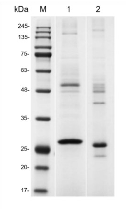
SDS-PAGE analysis of anti-CD3 monoclonal antibody in β mercaptoethanol-reduced (Lane 1) and non-reduced (Lane 2) conditions. Gel stained for 30 minutes with Coomassie Blue. As a result of different β-mercaptoethanol-reduced and non-reduced conditions, the target proteins migrate as about 25-35 kDa which is consistent with theoretical size.WB
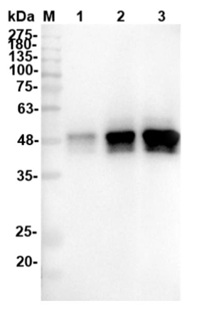
Western blot analysis of anti-CD3 monoclonal antibody was performed by loading human CD3 epsilon protein (Mouse IgG2a Fc Tag, 38.6 kDa) onto a 12% Tris-HCl polyacrylamide gel. Proteins were transferred to a CN membrane and blocked with 5% skim milk for one hour. Membranes were probed with anti-CD3 monoclonal antibody (2 ng/μL) and HRP mouse anti-His tag as a secondary antibody (1: 2000) in turn. Chemiluminescent detection was performed. Lane 1: Reducing antigen (0.1 μg) Lane 2: Reducing antigen (0.3 μg) Lane 3: Reducing antigen (0.6 μg) Lane 1: Reducing antigen (0.1 μg) Lane 2: Reducing antigen (0.3 μg) Lane 3: Reducing antigen (0.6 μg)| Product Name | Mouse Anti-CD3 Recombinant Antibody scFv Fragment |
|---|---|
| Antibody Type | Primary Antibodies |
| Product description | This anti-CD3 monoclonal antibody can be used in the treatment and optionally also prevention of diseases and disorders, such as autoimmune disorders, infectious diseases, and transplant rejection, that are susceptible to amelioration by binding to CD3. |
| Clonality | Monoclonal |
|---|---|
| Host Species | Mouse |
| Tested Applications | SDS-PAGEWB |
| WB:1:200-1:2000 |
|
| Species Reactivity | Human |
| Concentration | 1mg/ml |
| Purification | Affinity purified |
| Alternative Names | CD3 CD3 T-Cell Co-receptor CD3D CD3E CD3G |
|---|---|
| Molecular Weight(MW) | 25-35kDa |
| Function | In immunology, the CD3 (cluster of differentiation 3) T-cell co-receptor is a protein complex and is composed of four distinct chains. In mammals, the complex contains a CD3γ chain, a CD3δ chain, and two CD3ε chains. These chains associate with a molecule known as the T-cell receptor (TCR) and the ζ-chain to generate an activation signal in T lymphocytes. The TCR, ζ-chain, and CD3 molecules together comprise the TCR complex |
SDS-PAGE
SDS-PAGE analysis of anti-CD3 monoclonal antibody in β mercaptoethanol-reduced (Lane 1) and non-reduced (Lane 2) conditions. Gel stained for 30 minutes with Coomassie Blue. As a result of different β-mercaptoethanol-reduced and non-reduced conditions, the target proteins migrate as about 25-35 kDa which is consistent with theoretical size.
WB
Western blot analysis of anti-CD3 monoclonal antibody was performed by loading human CD3 epsilon protein (Mouse IgG2a Fc Tag, 38.6 kDa) onto a 12% Tris-HCl polyacrylamide gel. Proteins were transferred to a CN membrane and blocked with 5% skim milk for one hour. Membranes were probed with anti-CD3 monoclonal antibody (2 ng/μL) and HRP mouse anti-His tag as a secondary antibody (1: 2000) in turn. Chemiluminescent detection was performed. Lane 1: Reducing antigen (0.1 μg) Lane 2: Reducing antigen (0.3 μg) Lane 3: Reducing antigen (0.6 μg) Lane 1: Reducing antigen (0.1 μg) Lane 2: Reducing antigen (0.3 μg) Lane 3: Reducing antigen (0.6 μg)| Application Notes | WB:1:200-1:2000 |
|---|


