WB
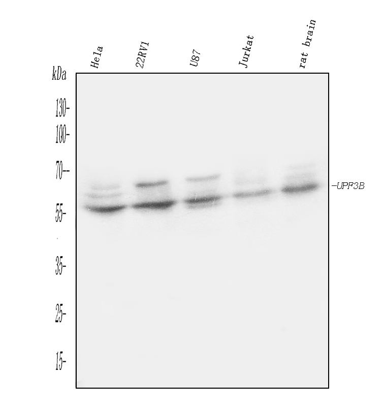
Western blot analysis of UPF3B using anti-UPF3B antibody. The sample well of each lane was loaded with 30 ug of sample under reducing conditions. Lane 1: Hela whole cell lysates, Lane 2: 22RV1 whole cell lysates, Lane 3: U87 whole cell lysates, Lane 4: JURKAT whole cell lysates, Lane 5: Rat brain tissue lysates. After electrophoresis, proteins were transferred to a membrane. Then the membrane was incubated with rabbit anti-UPF3B antigen affinity purified polyclonal antibody at a dilution of 1:1000 and probed with a goat anti-rabbit IgG-HRP secondary antibody. The signal is developed using ECL Plus Western Blotting Substrate.IHC
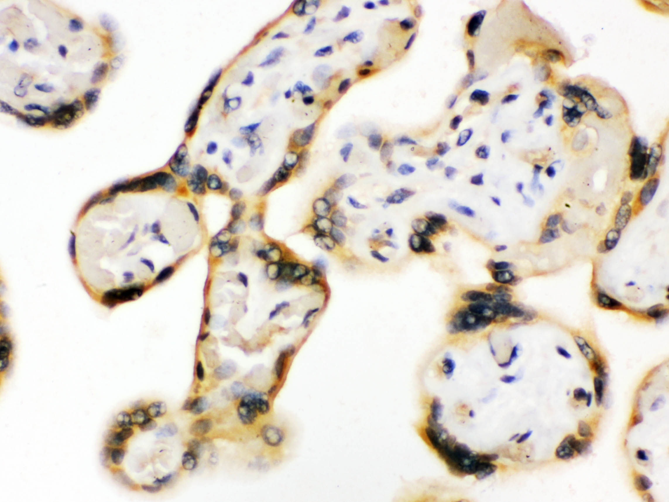
UPF3B was detected in a paraffin-embedded section of human placenta tissue. Biotinylated goat anti-rabbit IgG was used as secondary antibody. The tissue section was incubated with rabbit anti-UPF3B Antibody at a dilution of 1:200 and developed using Strepavidin-Biotin-Complex (SABC) with DAB as the chromogen.IHC
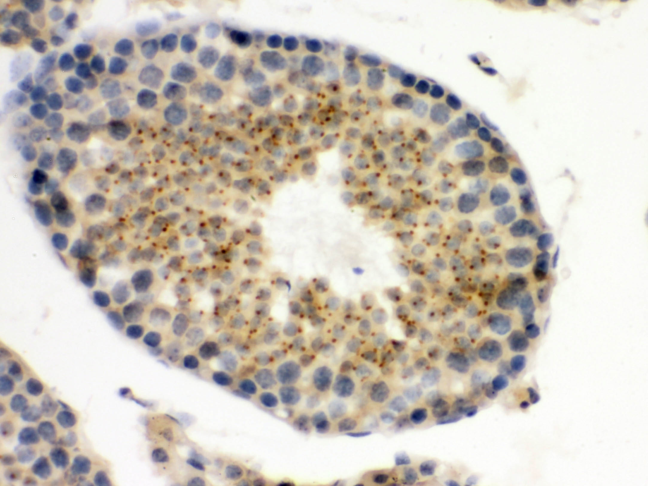
UPF3B was detected in a paraffin-embedded section of mouse testis tissue. Biotinylated goat anti-rabbit IgG was used as secondary antibody. The tissue section was incubated with rabbit anti-UPF3B Antibody at a dilution of 1:200 and developed using Strepavidin-Biotin-Complex (SABC) with DAB as the chromogen.IHC
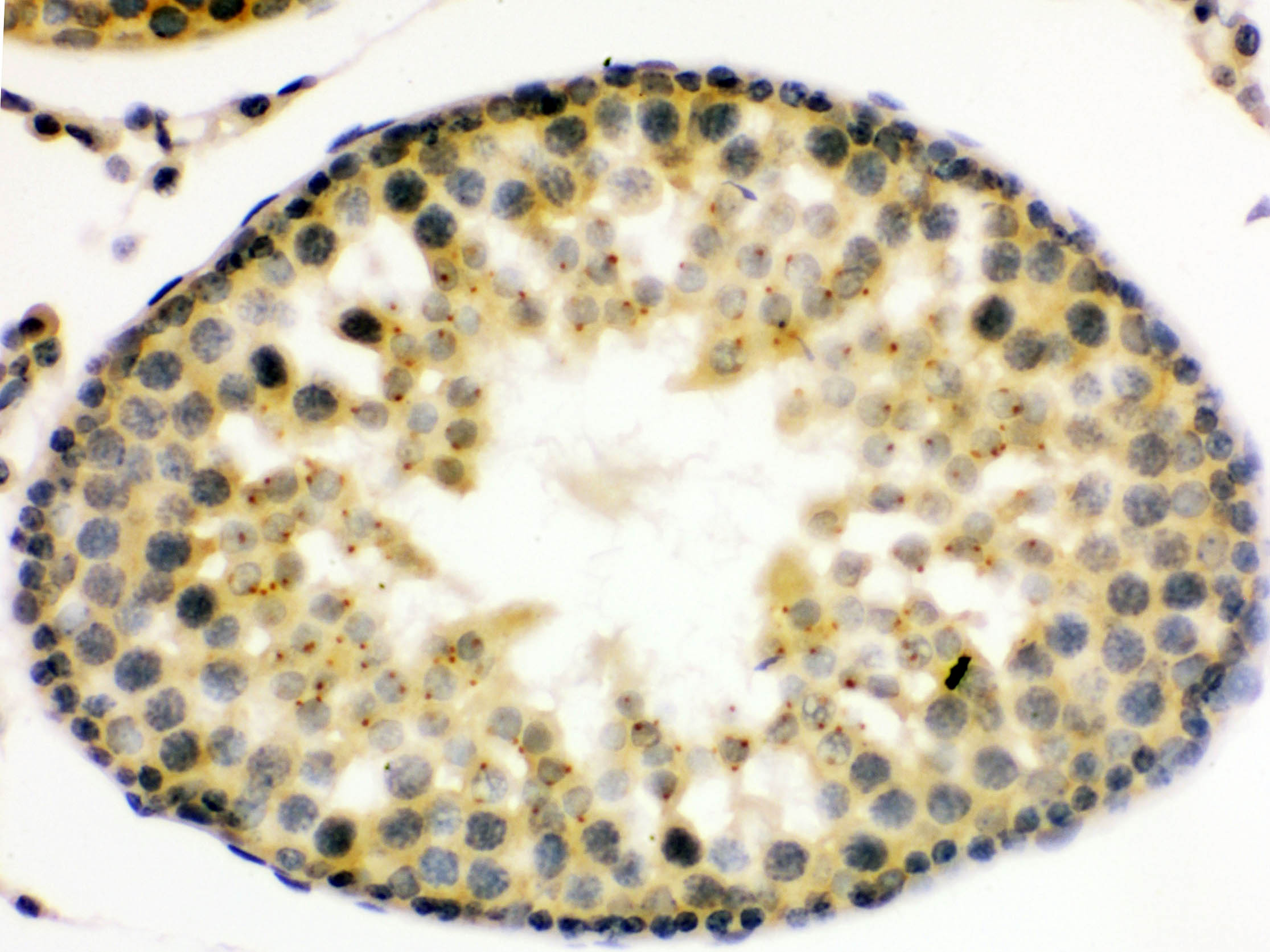
UPF3B was detected in a paraffin-embedded section of rat testis tissue. Biotinylated goat anti-rabbit IgG was used as secondary antibody. The tissue section was incubated with rabbit anti-UPF3B Antibody at a dilution of 1:200 and developed using Strepavidin-Biotin-Complex (SABC) with DAB as the chromogen.| Product Name | Rabbit polyclonal antibody to UPF3B |
|---|---|
| Antibody Type | Primary Antibodies |
| Immunogen | A synthetic peptide corresponding to a sequence at the C-terminus of human UPF3B /RENT3B |
| Clonality | Polyclonal |
|---|---|
| Isotype | IgG |
| Host Species | Rabbit |
| Tested Applications | IHCWB |
| WB:1:500-1:2000 IHC:1:50-1:400 |
|
| Species Reactivity | HumanMouseRat |
| Concentration | 0.5mg/ml |
| Purification | Affinity purified |
| Gene Symbol | UPF3B |
|---|---|
| Gene Synonyms | MRX62 MRX82 UPF3X HUPF3B MRXS14 RENT3B UPF3BP1 UPF3BP2 UPF3BP3 Upf3p-X |
| Gene Full Name | UPF3B regulator of nonsense mediated mRNA decay |
| Gene Summary | This gene encodes a protein that is part of a post-splicing multiprotein complex involved in both mRNA nuclear export and mRNA surveillance. The encoded protein is one of two functional homologs to yeast Upf3p. mRNA surveillance detects exported mRNAs with truncated open reading frames and initiates nonsense-mediated mRNA decay (NMD). When translation ends upstream from the last exon-exon junction, this triggers NMD to degrade mRNAs containing premature stop codons. This protein binds to the mRNA and remains bound after nuclear export, acting as a nucleocytoplasmic shuttling protein. It forms with Y14 a complex that binds specifically 20 nt upstream of exon-exon junctions. This gene is located on the long arm of chromosome X. Two splice variants encoding different isoforms have been found for this gene. [provided by RefSeq, Jul 2008] |
| Alternative Names | HUPF3B hUpf3p X MRXS14 RENT3B UPF3B UPF3X |
| Molecular Weight(MW) | 58 kDa |
| Cellular Localization | Cytoplasm,Nucleus. |
WB
Western blot analysis of UPF3B using anti-UPF3B antibody. The sample well of each lane was loaded with 30 ug of sample under reducing conditions. Lane 1: Hela whole cell lysates, Lane 2: 22RV1 whole cell lysates, Lane 3: U87 whole cell lysates, Lane 4: JURKAT whole cell lysates, Lane 5: Rat brain tissue lysates. After electrophoresis, proteins were transferred to a membrane. Then the membrane was incubated with rabbit anti-UPF3B antigen affinity purified polyclonal antibody at a dilution of 1:1000 and probed with a goat anti-rabbit IgG-HRP secondary antibody. The signal is developed using ECL Plus Western Blotting Substrate.
IHC
UPF3B was detected in a paraffin-embedded section of human placenta tissue. Biotinylated goat anti-rabbit IgG was used as secondary antibody. The tissue section was incubated with rabbit anti-UPF3B Antibody at a dilution of 1:200 and developed using Strepavidin-Biotin-Complex (SABC) with DAB as the chromogen.
IHC
UPF3B was detected in a paraffin-embedded section of mouse testis tissue. Biotinylated goat anti-rabbit IgG was used as secondary antibody. The tissue section was incubated with rabbit anti-UPF3B Antibody at a dilution of 1:200 and developed using Strepavidin-Biotin-Complex (SABC) with DAB as the chromogen.
IHC
UPF3B was detected in a paraffin-embedded section of rat testis tissue. Biotinylated goat anti-rabbit IgG was used as secondary antibody. The tissue section was incubated with rabbit anti-UPF3B Antibody at a dilution of 1:200 and developed using Strepavidin-Biotin-Complex (SABC) with DAB as the chromogen.| Application Notes | WB:1:500-1:2000 IHC:1:50-1:400 |
|---|
| Form | Liquid |
|---|---|
| Storage Instructions | 12 months from date of receipt, -20℃ as supplied. 6 months 2 to 8℃ after reconstitution. Avoid repeated freezing and thawing. |
| Storage Buffer | 500ug/ml antibody with PBS, 0.02% NaN3, 1 mg/ml BSA and 50% glycerol. |


