WB
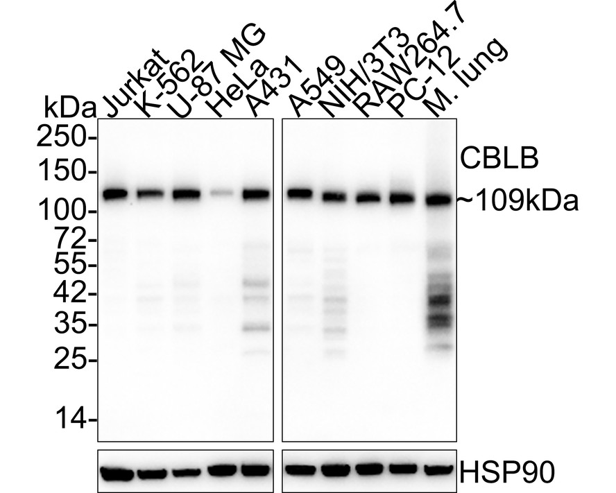
Western blot analysis of CBLB on different lysates with Rabbit anti-CBLB antibody at 1/2,000 dilution. Lane 1: Jurkat cell lysate (20 µg/Lane), Lane 2: K-562 cell lysate (20 µg/Lane), Lane 3: U-87 MG cell lysate (20 µg/Lane), Lane 4: HeLa cell lysate (20 µg/Lane), Lane 5: A431 cell lysate (20 µg/Lane), Lane 6: A549 cell lysate (20 µg/Lane), Lane 7: NIH/3T3 cell lysate (20 µg/Lane), Lane 8: RAW264.7 cell lysate (20 µg/Lane), Lane 9: PC-12 cell lysate (20 µg/Lane), Lane 10: Mouse lung tissue lysate (40 µg/Lane), Exposure time: 24 seconds; 4-20% SDS-PAGE gel. Proteins were transferred to a PVDF membrane and blocked with 5% NFDM/TBST for 1 hour at room temperature. The primary antibody at 1/2,000 dilution was used in 5% NFDM/TBST at 4℃ overnight. Goat Anti-Rabbit IgG - HRP Secondary Antibody at 1/50,000 dilution was used for 1 hour at room temperature.IHC
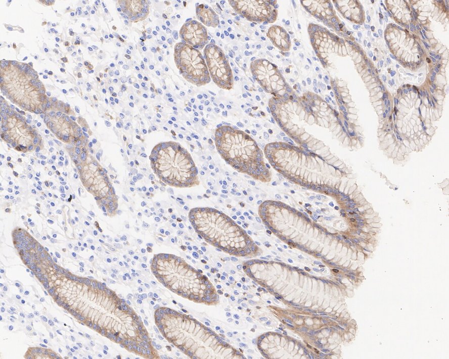
Immunohistochemical analysis of paraffin-embedded human stomach tissue with Rabbit anti-CBLB antibody at 1/200 dilution. The section was pre-treated using heat mediated antigen retrieval with Tris-EDTA buffer (pH 9.0) for 20 minutes. The tissues were blocked in 1% BSA for 20 minutes at room temperature, washed with ddH2O and PBS, and then probed with the primary antibody at 1/200 dilution for 1 hour at room temperature. The detection was performed using an HRP conjugated compact polymer system. DAB was used as the chromogen. Tissues were counterstained with hematoxylin and mounted with DPX.| Product Name | CBLB Recombinant Rabbit Monoclonal Antibody |
|---|---|
| Antibody Type | Primary Antibodies |
| Immunogen | Recombinant protein within human CBLB aa 400-982 / 982. |
| Clonality | Monoclonal |
|---|---|
| Isotype | IgG |
| Host Species | Rabbit |
| Tested Applications | IHCWB |
| WB:1:2000 IHC:1:200 |
|
| Species Reactivity | HumanMouseRat |
| Concentration | 1mg/ml |
| Purification | Protein A |
| Gene Symbol | CBLB |
|---|---|
| Gene Synonyms | Cbl-b RNF56 ADMIO3 Nbla00127 |
| Gene Full Name | Cbl proto-oncogene B |
| Gene Summary | This gene encodes an E3 ubiquitin-protein ligase which promotes proteosome-mediated protein degradation by transferring ubiquitin from an E2 ubiquitin-conjugating enzyme to a substrate. The encoded protein is involved in the regulation of immune response by limiting T-cell receptor, B-cell receptor, and high affinity immunoglobulin epsilon receptor activation. Studies in mouse suggest that this gene is involved in antifungal host defense and that its inhibition leads to increased fungal killing. Manipulation of this gene may be beneficial in implementing immunotherapies for a variety of conditions, including cancer, autoimmune diseases, allergies, and infections. [provided by RefSeq, Sep 2017] |
| Alternative Names | AI429560 antibody AI851073 antibody Cas Br M (murine) ecotropic retroviral transforming sequence b antibody Casitas B lineage lymphoma b antibody Casitas B lineage lymphoma proto oncogene b antibody Casitas B-lineage lymphoma proto-oncogene b antibody Cbl b antibody CBLB antibody CBLB_HUMAN antibody DKFZp686J10223 antibody AI429560 antibody AI851073 antibody Cas Br M (murine) ecotropic retroviral transforming sequence b antibody Casitas B lineage lymphoma b antibody Casitas B lineage lymphoma proto oncogene b antibody Casitas B-lineage lymphoma proto-oncogene b antibody Cbl b antibody CBLB antibody CBLB_HUMAN antibody DKFZp686J10223 antibody DKFZp779A0729 antibody DKFZp779F1443 antibody E3 ubiquitin protein ligase CBL B antibody E3 ubiquitin-protein ligase CBL-B antibody EC 6.3.2.- antibody FLJ36865 antibody FLJ41152 antibody Nbla00127 antibody RING finger protein 56 antibody RNF56 antibody SH3 binding protein CBL B antibody SH3-binding protein CBL-B antibody Signal transduction protein CBL B antibody Signal transduction protein CBL-B antibody |
| Molecular Weight(MW) | 109kDa |
| Cellular Localization | Cytoplasm. |
WB
Western blot analysis of CBLB on different lysates with Rabbit anti-CBLB antibody at 1/2,000 dilution. Lane 1: Jurkat cell lysate (20 µg/Lane), Lane 2: K-562 cell lysate (20 µg/Lane), Lane 3: U-87 MG cell lysate (20 µg/Lane), Lane 4: HeLa cell lysate (20 µg/Lane), Lane 5: A431 cell lysate (20 µg/Lane), Lane 6: A549 cell lysate (20 µg/Lane), Lane 7: NIH/3T3 cell lysate (20 µg/Lane), Lane 8: RAW264.7 cell lysate (20 µg/Lane), Lane 9: PC-12 cell lysate (20 µg/Lane), Lane 10: Mouse lung tissue lysate (40 µg/Lane), Exposure time: 24 seconds; 4-20% SDS-PAGE gel. Proteins were transferred to a PVDF membrane and blocked with 5% NFDM/TBST for 1 hour at room temperature. The primary antibody at 1/2,000 dilution was used in 5% NFDM/TBST at 4℃ overnight. Goat Anti-Rabbit IgG - HRP Secondary Antibody at 1/50,000 dilution was used for 1 hour at room temperature.
IHC
Immunohistochemical analysis of paraffin-embedded human stomach tissue with Rabbit anti-CBLB antibody at 1/200 dilution. The section was pre-treated using heat mediated antigen retrieval with Tris-EDTA buffer (pH 9.0) for 20 minutes. The tissues were blocked in 1% BSA for 20 minutes at room temperature, washed with ddH2O and PBS, and then probed with the primary antibody at 1/200 dilution for 1 hour at room temperature. The detection was performed using an HRP conjugated compact polymer system. DAB was used as the chromogen. Tissues were counterstained with hematoxylin and mounted with DPX.| Application Notes | WB:1:2000 IHC:1:200 |
|---|
| Form | Liquid |
|---|---|
| Storage Instructions | Store at +4℃ after thawing. Aliquot store at -20℃. Avoid repeated freeze / thaw cycles. |
| Storage Buffer | PBS (pH7.4), 0.1% BSA, 40% Glycerol. Preservative: 0.05% Sodium Azide. |


