WB
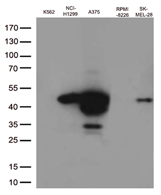
Western blot analysis of extracts (35ug) from 5 cell lines lysates by using anti-MAGEA4 monoclonal antibody (1:500).IHC
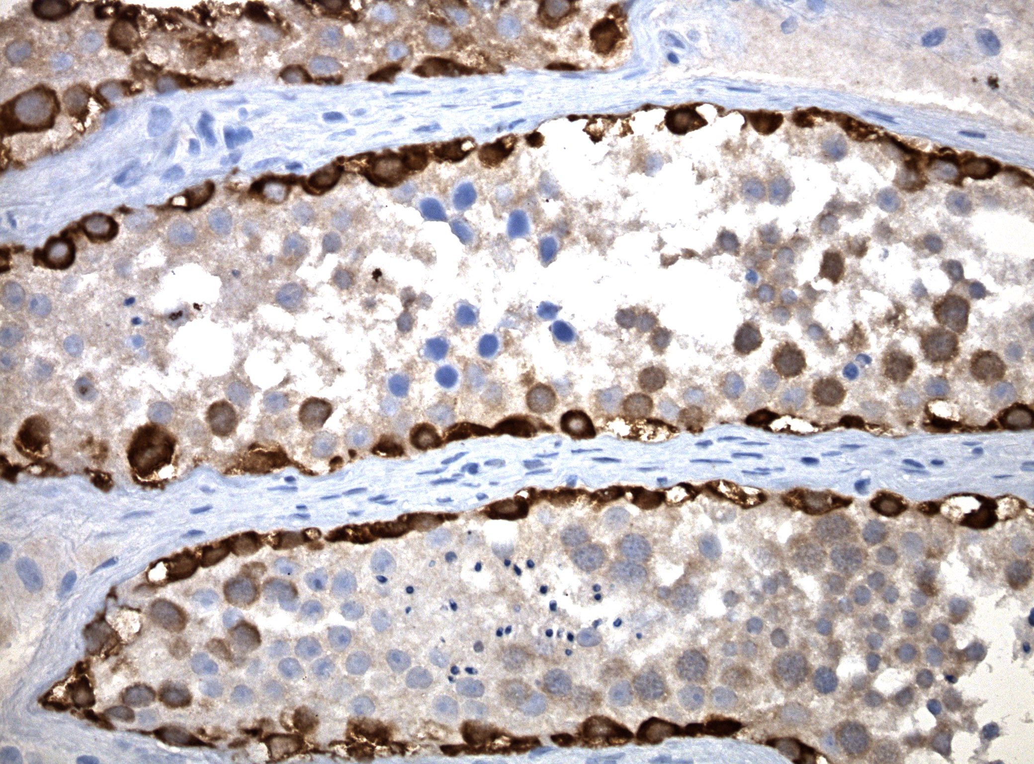
Immunohistochemical staining of paraffin-embedded testis using anti-MAGE-A4 mouse monoclonal antibody. HIER TEE buffer pH9 at 110C for 10 min, anti-MAGE-A4 diluted to 1:50. Detection was done with Polink1 Broad Mouse and Rabbit with DAB Kit. Image 40x magnification.IHC
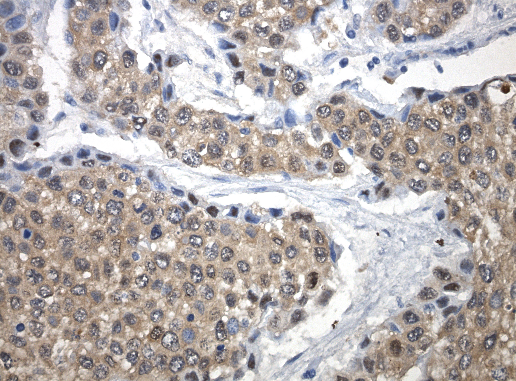
Immunohistochemical staining of paraffin-embedded bladder cancer using anti-MAGE-A4 mouse monoclonal antibody. HIER TEE buffer pH9 at 110C for 10 min, anti-MAGE-A4 diluted to 1:100. Detection was done with Polink1 Broad Mouse and Rabbit with DAB Kit. Image 40x magnification.FC
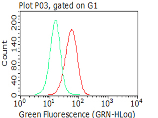
Flow cytometric analysis of fixed and permeabilized NCI-H1299 cells, using anti-MAGEA4 antibody, compared to an isotype control (green) (1:100).| Product Name | MAGEA4 Mouse Monoclonal Antibody |
|---|---|
| Antibody Type | Primary Antibodies |
| Immunogen | Full length human recombinant protein of human MAGEA4(NP_001011550) produced in HEK293T cell. |
| Clonality | monoclonal |
|---|---|
| Isotype | IgG2a |
| Host Species | Mouse |
| Tested Applications | FCIHCWB |
| WB:1:500-1:2000 IHC:1:50-1:200 FC:1:100 |
|
| Species Reactivity | Human |
| Concentration | 1mg/ml |
| Purification | Protein A/G |
| Gene Symbol | MAGEA4 |
|---|---|
| Gene Synonyms | CT1.4 MAGE4 MAGE4A MAGE4B MAGE-41 MAGE-X2 |
| Gene Full Name | MAGE family member A4 |
| Gene Summary | This gene is a member of the MAGEA gene family. The members of this family encode proteins with 50 to 80% sequence identity to each other. The promoters and first exons of the MAGEA genes show considerable variability, suggesting that the existence of this gene family enables the same function to be expressed under different transcriptional controls. The MAGEA genes are clustered at chromosomal location Xq28. They have been implicated in some hereditary disorders, such as dyskeratosis congenita. Several variants encoding the same protein have been found for this gene. [provided by RefSeq, Aug 2020] |
| Molecular Weight(MW) | 35kDa |
| Cellular Localization | nucleus. |
WB
Western blot analysis of extracts (35ug) from 5 cell lines lysates by using anti-MAGEA4 monoclonal antibody (1:500).
IHC
Immunohistochemical staining of paraffin-embedded testis using anti-MAGE-A4 mouse monoclonal antibody. HIER TEE buffer pH9 at 110C for 10 min, anti-MAGE-A4 diluted to 1:50. Detection was done with Polink1 Broad Mouse and Rabbit with DAB Kit. Image 40x magnification.
IHC
Immunohistochemical staining of paraffin-embedded bladder cancer using anti-MAGE-A4 mouse monoclonal antibody. HIER TEE buffer pH9 at 110C for 10 min, anti-MAGE-A4 diluted to 1:100. Detection was done with Polink1 Broad Mouse and Rabbit with DAB Kit. Image 40x magnification.
FC
Flow cytometric analysis of fixed and permeabilized NCI-H1299 cells, using anti-MAGEA4 antibody, compared to an isotype control (green) (1:100).| Application Notes | WB:1:500-1:2000 IHC:1:50-1:200 FC:1:100 |
|---|
| Form | Liquid |
|---|---|
| Storage Instructions | Store at -20°C as received. |
| Storage Buffer | PBS (pH 7.3) containing 1% BSA, 50% glycerol and 0.02% sodium azide. |


