
IHC
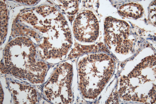
IHC image of MAGEA4 Monoclonal Antibody diluted at 1:200 and staining in paraffin-embedded human testis tissue performed on a Leica BondTM system. After dewaxing and hydration, antigen retrieval was mediated by high pressure in a citrate buffer (pH 6.0). Section was blocked with 10% normal goat serum 30min at RT. Then primary antibody (1% BSA) was incubated at 4°C overnight. The primary is detected by a Goat anti-mouse polymer IgG labeled by HRP and visualized using 0.05% DAB.IHC
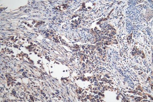
IHC image of MAGEA4 Monoclonal Antibody diluted at 1:200 and staining in paraffin-embedded human bladder cancer performed on a Leica BondTM system. After dewaxing and hydration, antigen retrieval was mediated by high pressure in a citrate buffer (pH 6.0). Section was blocked with 10% normal goat serum 30min at RT. Then primary antibody (1% BSA) was incubated at 4°C overnight. The primary is detected by a Goat anti-mouse polymer IgG labeled by HRP and visualized using 0.05% DAB.IHC
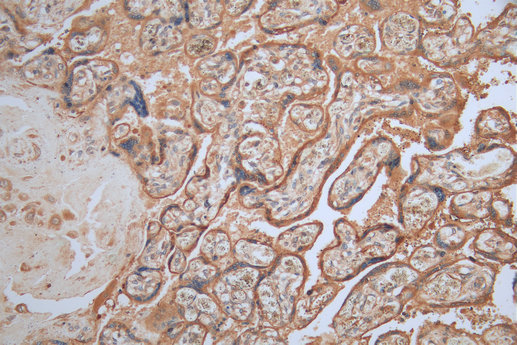
IHC image of MAGEA4 Monoclonal Antibody diluted at 1:200 and staining in paraffin-embedded human placenta tissue performed on a Leica BondTM system. After dewaxing and hydration, antigen retrieval was mediated by high pressure in a citrate buffer (pH 6.0). Section was blocked with 10% normal goat serum 30min at RT. Then primary antibody (1% BSA) was incubated at 4°C overnight. The primary is detected by a Goat anti-mouse polymer IgG labeled by HRP and visualized using 0.05% DAB.IHC
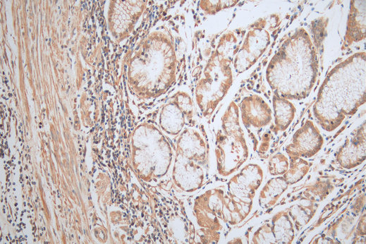
IHC image of MAGEA4 Monoclonal Antibody diluted at 1:200 and staining in paraffin-embedded human gastric cancer tissue performed on a Leica BondTM system. After dewaxing and hydration, antigen retrieval was mediated by high pressure in a citrate buffer (pH 6.0). Section was blocked with 10% normal goat serum 30min at RT. Then primary antibody (1% BSA) was incubated at 4°C overnight. The primary is detected by a Goat anti-mouse polymer IgG labeled by HRP and visualized using 0.05% DAB.FC
Overlay Peak curve showing A375 cells stained with MAGEA4 Monoclonal Antibody (red line) at 1:100. Then 10% normal goat serum was Incubated to block non-specific protein-protein interactions followed by the antibody (1µg/1*106cells) for 45 min at 4°C. The secondary antibody used was FITC-conjugated Goat Anti-Mouse IgG(H+L) at 1/200 dilution for 35 min at 4°C. Isotype control antibody (green line) was mouse IgG1 (1µg/1*106cells) used under the same conditions. Acquisition of >10,000 events was performed.| Product Name | Mouse anti-MAGEA4 Monoclonal antibody |
|---|---|
| Antibody Type | Primary Antibodies |
| Immunogen | Recombinant Human Melanoma-associated antigen 4 protein (1-317AA). |
| Clonality | Monoclonal |
|---|---|
| Isotype | IgG1 |
| Host Species | Mouse |
| Tested Applications | FCIHC |
| IHC:1:200-1:500 FC:1:50-1:200 |
|
| Species Reactivity | Human |
| Concentration | 1mg/ml |
| Purification | Protein G |
| Gene Symbol | MAGEA4 |
|---|---|
| Gene Synonyms | CT1.4 MAGE4 MAGE4A MAGE4B MAGE-41 MAGE-X2 |
| Gene Full Name | MAGE family member A4 |
| Gene Summary | This gene is a member of the MAGEA gene family. The members of this family encode proteins with 50 to 80% sequence identity to each other. The promoters and first exons of the MAGEA genes show considerable variability, suggesting that the existence of this gene family enables the same function to be expressed under different transcriptional controls. The MAGEA genes are clustered at chromosomal location Xq28. They have been implicated in some hereditary disorders, such as dyskeratosis congenita. Several variants encoding the same protein have been found for this gene. [provided by RefSeq, Aug 2020] |
| Alternative Names | Cancer/testis antigen 1.4 antibody CT1.4 antibody MAGA4_HUMAN antibody MAGE 4 antibody MAGE 4 antigen antibody MAGE 41 antibody MAGE 41 antigen antibody MAGE 4A antibody MAGE 4B antibody MAGE X2 antibody MAGE X2 antigen antibody MAGE-4 antigen antibody MAGE-41 antigen antibody MAGE-X2 antigen antibody MAGE4 antibody MAGE41 antibody MAGE4A antibody MAGE4B antibody MAGEA 4 antibody MAGEA4 antibody Melanoma antigen family A 4 antibody Melanoma associated antigen 4 antibody Melanoma-associated antigen 4 antibody MGC21336 antibody |
IHC
IHC image of MAGEA4 Monoclonal Antibody diluted at 1:200 and staining in paraffin-embedded human testis tissue performed on a Leica BondTM system. After dewaxing and hydration, antigen retrieval was mediated by high pressure in a citrate buffer (pH 6.0). Section was blocked with 10% normal goat serum 30min at RT. Then primary antibody (1% BSA) was incubated at 4°C overnight. The primary is detected by a Goat anti-mouse polymer IgG labeled by HRP and visualized using 0.05% DAB.
IHC
IHC image of MAGEA4 Monoclonal Antibody diluted at 1:200 and staining in paraffin-embedded human bladder cancer performed on a Leica BondTM system. After dewaxing and hydration, antigen retrieval was mediated by high pressure in a citrate buffer (pH 6.0). Section was blocked with 10% normal goat serum 30min at RT. Then primary antibody (1% BSA) was incubated at 4°C overnight. The primary is detected by a Goat anti-mouse polymer IgG labeled by HRP and visualized using 0.05% DAB.
IHC
IHC image of MAGEA4 Monoclonal Antibody diluted at 1:200 and staining in paraffin-embedded human placenta tissue performed on a Leica BondTM system. After dewaxing and hydration, antigen retrieval was mediated by high pressure in a citrate buffer (pH 6.0). Section was blocked with 10% normal goat serum 30min at RT. Then primary antibody (1% BSA) was incubated at 4°C overnight. The primary is detected by a Goat anti-mouse polymer IgG labeled by HRP and visualized using 0.05% DAB.
IHC
IHC image of MAGEA4 Monoclonal Antibody diluted at 1:200 and staining in paraffin-embedded human gastric cancer tissue performed on a Leica BondTM system. After dewaxing and hydration, antigen retrieval was mediated by high pressure in a citrate buffer (pH 6.0). Section was blocked with 10% normal goat serum 30min at RT. Then primary antibody (1% BSA) was incubated at 4°C overnight. The primary is detected by a Goat anti-mouse polymer IgG labeled by HRP and visualized using 0.05% DAB.
FC
Overlay Peak curve showing A375 cells stained with MAGEA4 Monoclonal Antibody (red line) at 1:100. Then 10% normal goat serum was Incubated to block non-specific protein-protein interactions followed by the antibody (1µg/1*106cells) for 45 min at 4°C. The secondary antibody used was FITC-conjugated Goat Anti-Mouse IgG(H+L) at 1/200 dilution for 35 min at 4°C. Isotype control antibody (green line) was mouse IgG1 (1µg/1*106cells) used under the same conditions. Acquisition of >10,000 events was performed.| Application Notes | IHC:1:200-1:500 FC:1:50-1:200 |
|---|
| Form | Liquid |
|---|---|
| Storage Instructions | Upon receipt, store at -20°C or -80°C. Avoid repeated freeze. |
| Storage Buffer | Preservative: 0.03% Proclin 300,Constituents: 50% Glycerol, 0.01M PBS, pH 7.4 |


