WB
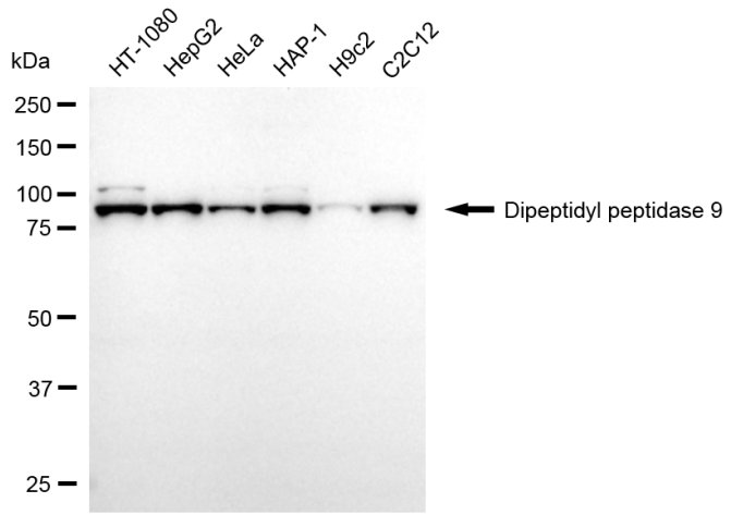
Western blotting analysis using dipeptidyl peptidase 9 antibody. Total cell lysates (30 μg) from various cell lines were loaded and separated by SDS-PAGE. The blot was incubated with dipeptidyl peptidase 9 antibody and HRP-conjugated goat anti-mouse secondary antibody respectively.ICC/IF
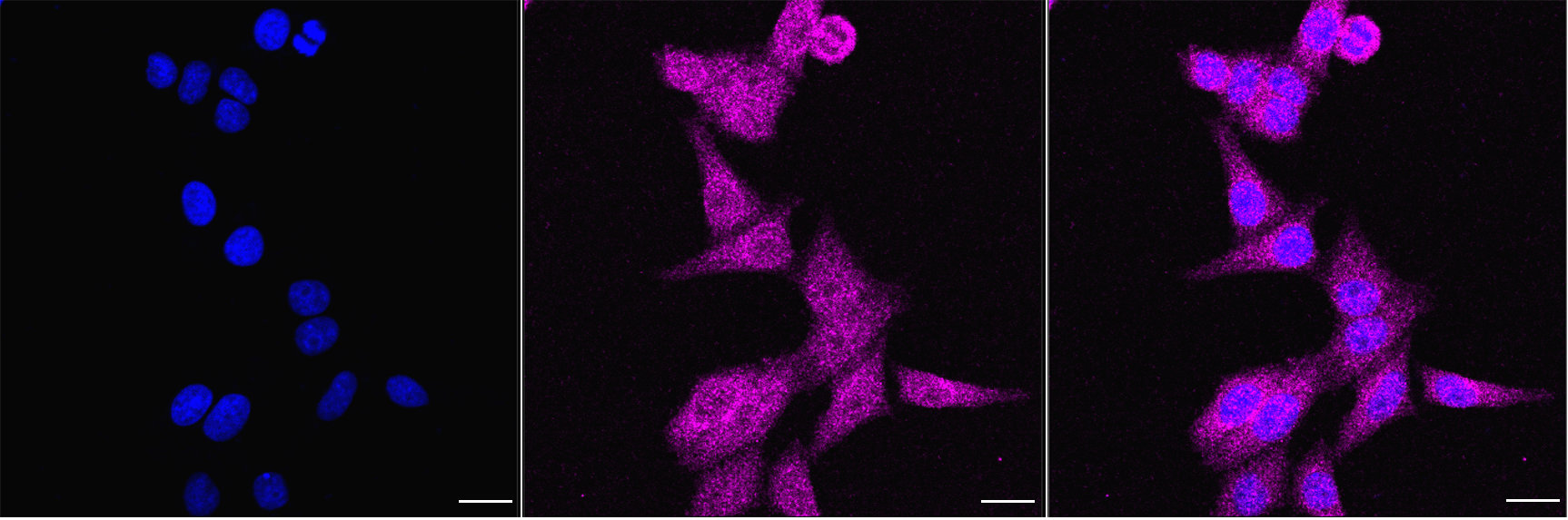
Immunocytochemical staining of HepG2 cells with Dipeptidyl peptidase 9 antibody. Nuclei were stained blue with DAPI; Dipeptidyl peptidase 9 was stained magenta with Alexa Fluor® 647. Images were taken using Leica stellaris 5. Protein abundance based on laser Intensity and smart gain: Low. Scale bar, 20 μm.FC
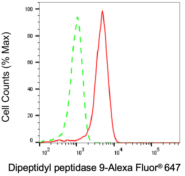
Flow cytometric analysis of Dipeptidyl peptidase 9 expression in HepG2 cells using Dipeptidyl peptidase 9 antibody. Green, isotype control; red, Dipeptidyl peptidase 9.| Product Name | Mouse anti-DPP9 Monoclonal antibody |
|---|---|
| Antibody Type | Primary Antibodies |
| Immunogen | Recombinant Human DPP9 protein |
| Clonality | Monoclonal |
|---|---|
| Isotype | IgG1 |
| Host Species | Mouse |
| Tested Applications | FCICC/IFWB |
| WB:1:2500-1:5000 ICC/IF:1:100-1:1000 FC:1:100-1:1000 |
|
| Species Reactivity | HumanMouseRat |
| Concentration | 1mg/ml |
| Purification | Affinity purified |
| Gene Symbol | DPP9 |
|---|---|
| Gene Synonyms | DP9 DPLP9 DPRP2 HATIS DPP IX DPRP-2 IMD111 |
| Gene Full Name | dipeptidyl peptidase 9 |
| Gene Summary | This gene encodes a protein that is a member of the S9B family in clan SC of the serine proteases. The protein has been shown to have post-proline dipeptidyl aminopeptidase activity, cleaving Xaa-Pro dipeptides from the N-termini of proteins. Although the activity of this protein is similar to that of dipeptidyl peptidase 4 (DPP4), it does not appear to be membrane bound. In general, dipeptidyl peptidases appear to be involved in the regulation of the activity of their substrates and have been linked to a variety of diseases including type 2 diabetes, obesity and cancer. Several transcript variants of this gene have been described but not fully characterized. [provided by RefSeq, Jul 2008] |
| Alternative Names | DPP9 Dipeptidyl Peptidase 9 Dipeptidyl Peptidase-Like Protein 9 Dipeptidyl Peptidase IX DPP IX DPRP-2 DPLP9 DPRP2 DP9 Dipeptidyl Peptidase IV-Related Protein-2 Dipeptidyl Peptidase IV-Related Protein 2 Dipeptidylpeptidase 9 EC 3.4.14.5 IMD111 HATIS |
| Molecular Weight(MW) | 98 kDa |
| Cellular Localization | Cytoplasm, cytosol, Nucleus. |
WB
Western blotting analysis using dipeptidyl peptidase 9 antibody. Total cell lysates (30 μg) from various cell lines were loaded and separated by SDS-PAGE. The blot was incubated with dipeptidyl peptidase 9 antibody and HRP-conjugated goat anti-mouse secondary antibody respectively.
ICC/IF
Immunocytochemical staining of HepG2 cells with Dipeptidyl peptidase 9 antibody. Nuclei were stained blue with DAPI; Dipeptidyl peptidase 9 was stained magenta with Alexa Fluor® 647. Images were taken using Leica stellaris 5. Protein abundance based on laser Intensity and smart gain: Low. Scale bar, 20 μm.
FC
Flow cytometric analysis of Dipeptidyl peptidase 9 expression in HepG2 cells using Dipeptidyl peptidase 9 antibody. Green, isotype control; red, Dipeptidyl peptidase 9.| Application Notes | WB:1:2500-1:5000 ICC/IF:1:100-1:1000 FC:1:100-1:1000 |
|---|
| Form | Liquid |
|---|---|
| Storage Instructions | Upon receipt, store at -20°C or -80°C. Avoid repeated freeze. |
| Storage Buffer | 0.03% Proclin 300 Constituents: 50% Glycerol, 0.01M PBS, PH 7.4 |


