WB
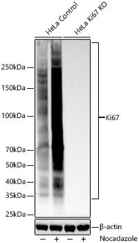
Western blot analysis of lysates from wild type (WT) and Ki67 knockout (KO) HeLa cells using Ki67 Rabbit mAb at 1:5000 dilution incubated overnight at 4℃. HeLa cells were treated with Nocadazole (1 μg/mL) at 37℃ for 16 hours Secondary antibody: HRP-conjugated Goat anti-Rabbit IgG (H+L)at 1:10000 dilution. Lysates/proteins: 25 μg per lane. Blocking buffer: 3% nonfat dry milk in TBST. Detection: ECL Basic Kit. Exposure time: 45s.IHC
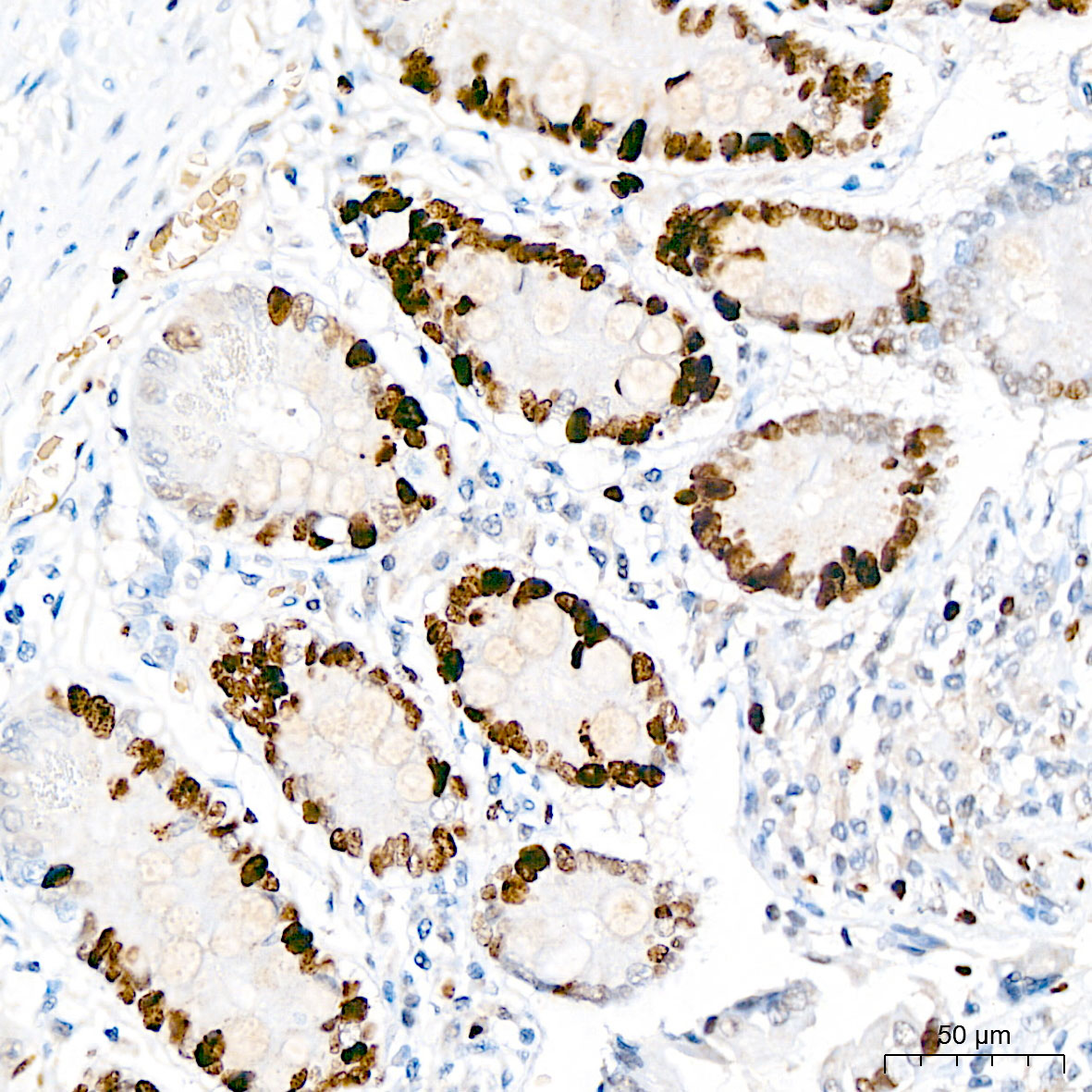
Immunohistochemistry analysis of paraffin embedded Human small intestine tissue using Ki67 Rabbit mAb at a dilution of 1:500 (40x lens). High pressure antigen retrieval performed with 0.01M Citrate Buffer (pH 6.0) prior to IHC staining.IHC
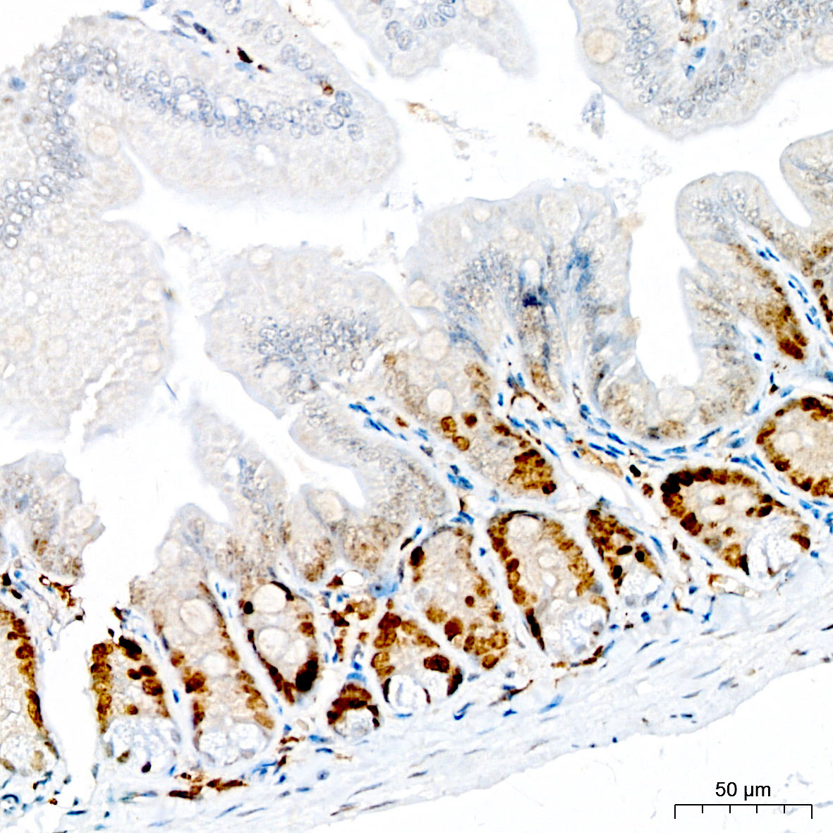
Immunohistochemistry analysis of paraffin embedded Mouse intestine tissue using Ki67 Rabbit mAb at a dilution of 1:500 (40x lens). High pressure antigen retrieval performed with 0.01M Citrate Buffer (pH 6.0) prior to IHC staining.IHC
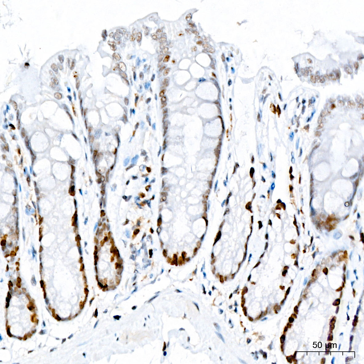
Immunohistochemistry analysis of paraffin embedded Rat colon tissue using Ki67 Rabbit mAb at a dilution of 1:500 (40x lens). High pressure antigen retrieval performed with 0.01M Citrate Buffer (pH 6.0) prior to IHC staining.ICC/IF
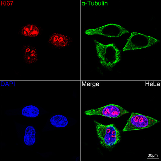
Confocal imaging of HeLa cells using Ki67 Rabbit mAb (dilution 1:100)(Red) followed by a further incubation with Cy3-conjugated Goat Anti-Rabbit IgG (H+L) (dilution 1:500) (Red). The cells were counterstained with α-Tubulin Mouse mAb (dilution 1:400) followed by incubation with Omnimabs® 488 Goat Anti-Mouse IgG(H&L) (dilution 1:500) (Green). DAPI was used for nuclear staining (Blue). Objective: 100x.mIHC
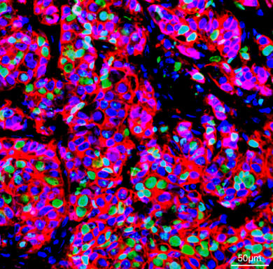
The multiplex IHC analysis on paraffin embedded Human breast cancer tissue using the following specific primary antibodies and tyramide signal amplification (TSA) reagents: Ki67 Rabbit mAb (1:500) with TSA-TYR-520 (Green), HER2/ErbB2 Rabbit mAb (1:200) with TSA-TYR-570 (Red), and Androgen Receptor Rabbit mAb (1:400) with TSA-TYR-690 (Magenta). DAPI (Blue) was used for nuclear staining. Prior to multiplex IHC staining, high-pressure antigen retrieval was performed using 0.01M citrate buffer at pH 6.0. The analysis was completed using a 20x objective lens.| Product Name | Ki67 Rabbit mAb |
|---|---|
| Antibody Type | Primary Antibodies |
| Immunogen | Synthetic peptide. |
| Clonality | Monoclonal |
|---|---|
| Isotype | IgG |
| Host Species | Rabbit |
| Tested Applications | ICC/IFIF-PIHCWB |
| WB:1:2000-1:10000 IHC:1:200-1:800 ICC/IF:1:50-1:200 mIHC:1:200-1:800 |
|
| Species Reactivity | HumanMouseRat |
| Concentration | 1mg/ml |
| Purification | Affinity purified |
| Gene Symbol | MKI67 |
|---|---|
| Gene Synonyms | KIA MIB- MIB-1 PPP1R105 |
| Gene Full Name | marker of proliferation Ki-67 |
| Gene Summary | Enables RNA binding activity and molecular condensate scaffold activity. Involved in chromosome segregation and regulation of mitotic nuclear division. Located in chromosome; nucleolus; and nucleoplasm. Is active in condensed chromosome. Implicated in several diseases, including Crohn's disease; colorectal cancer; endocrine gland cancer (multiple); graft-versus-host disease; and human immunodeficiency virus infectious disease. Biomarker of several diseases, including Barrett's esophagus; autoimmune disease of musculoskeletal system (multiple); endocrine gland cancer (multiple); gastrointestinal system cancer (multiple); and lung cancer (multiple). [provided by Alliance of Genome Resources, Jul 2025] |
| Alternative Names | KIA MIB- MIB-1 PPP1R105 Ki67 |
| Molecular Weight(MW) | 359 kDa |
| Cellular Localization | Chromosome, Nucleus, nucleolus. |
WB
Western blot analysis of lysates from wild type (WT) and Ki67 knockout (KO) HeLa cells using Ki67 Rabbit mAb at 1:5000 dilution incubated overnight at 4℃. HeLa cells were treated with Nocadazole (1 μg/mL) at 37℃ for 16 hours Secondary antibody: HRP-conjugated Goat anti-Rabbit IgG (H+L)at 1:10000 dilution. Lysates/proteins: 25 μg per lane. Blocking buffer: 3% nonfat dry milk in TBST. Detection: ECL Basic Kit. Exposure time: 45s.
IHC
Immunohistochemistry analysis of paraffin embedded Human small intestine tissue using Ki67 Rabbit mAb at a dilution of 1:500 (40x lens). High pressure antigen retrieval performed with 0.01M Citrate Buffer (pH 6.0) prior to IHC staining.
IHC
Immunohistochemistry analysis of paraffin embedded Mouse intestine tissue using Ki67 Rabbit mAb at a dilution of 1:500 (40x lens). High pressure antigen retrieval performed with 0.01M Citrate Buffer (pH 6.0) prior to IHC staining.
IHC
Immunohistochemistry analysis of paraffin embedded Rat colon tissue using Ki67 Rabbit mAb at a dilution of 1:500 (40x lens). High pressure antigen retrieval performed with 0.01M Citrate Buffer (pH 6.0) prior to IHC staining.
ICC/IF
Confocal imaging of HeLa cells using Ki67 Rabbit mAb (dilution 1:100)(Red) followed by a further incubation with Cy3-conjugated Goat Anti-Rabbit IgG (H+L) (dilution 1:500) (Red). The cells were counterstained with α-Tubulin Mouse mAb (dilution 1:400) followed by incubation with Omnimabs® 488 Goat Anti-Mouse IgG(H&L) (dilution 1:500) (Green). DAPI was used for nuclear staining (Blue). Objective: 100x.
mIHC
The multiplex IHC analysis on paraffin embedded Human breast cancer tissue using the following specific primary antibodies and tyramide signal amplification (TSA) reagents: Ki67 Rabbit mAb (1:500) with TSA-TYR-520 (Green), HER2/ErbB2 Rabbit mAb (1:200) with TSA-TYR-570 (Red), and Androgen Receptor Rabbit mAb (1:400) with TSA-TYR-690 (Magenta). DAPI (Blue) was used for nuclear staining. Prior to multiplex IHC staining, high-pressure antigen retrieval was performed using 0.01M citrate buffer at pH 6.0. The analysis was completed using a 20x objective lens.| Application Notes | WB:1:2000-1:10000 IHC:1:200-1:800 ICC/IF:1:50-1:200 mIHC:1:200-1:800 |
|---|
| Form | Liquid |
|---|---|
| Storage Instructions | Store at -20℃. Avoid freeze / thaw cycles. |
| Storage Buffer | Buffer: PBS with 0.05% proclin300, 0.05% BSA, 50% glycerol, pH7.3. |


