WB
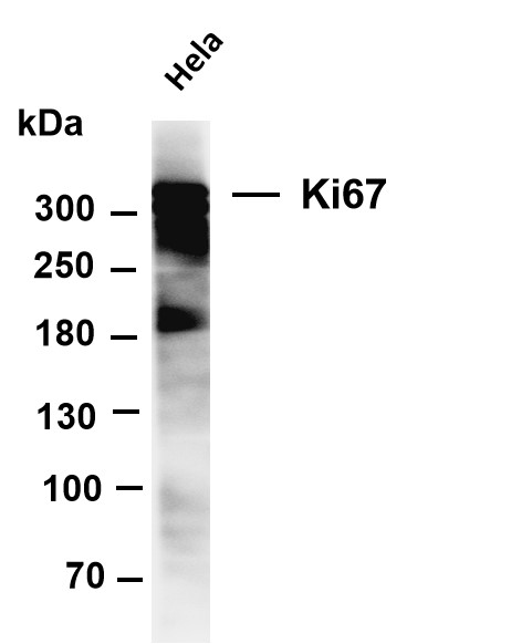
Various whole cell lysates were separated by 4-8% SDS-PAGE, and the membrane was blotted with anti-Ki67 antibody. The HRP-conjugated Goat anti-Rabbit IgG(H + L) antibody was used to detect the antibody. Lane 1: Hela.IHC
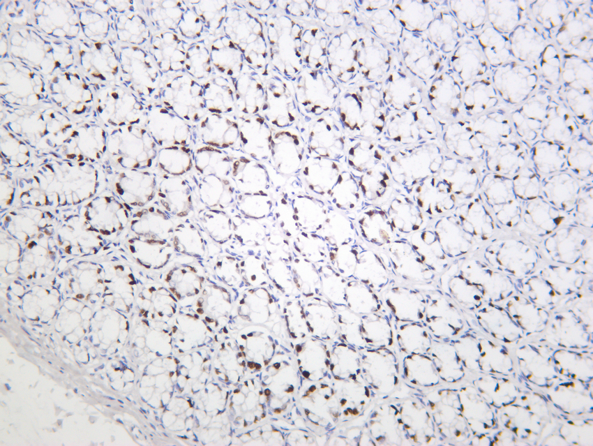
Rat colon was stained with Anti-Ki67 rabbit antibody.IHC
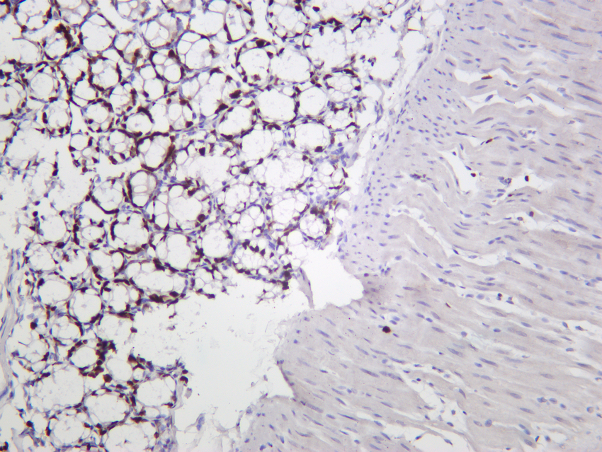
Mouse colon was stained with Anti-Ki67 rabbit antibody.IHC
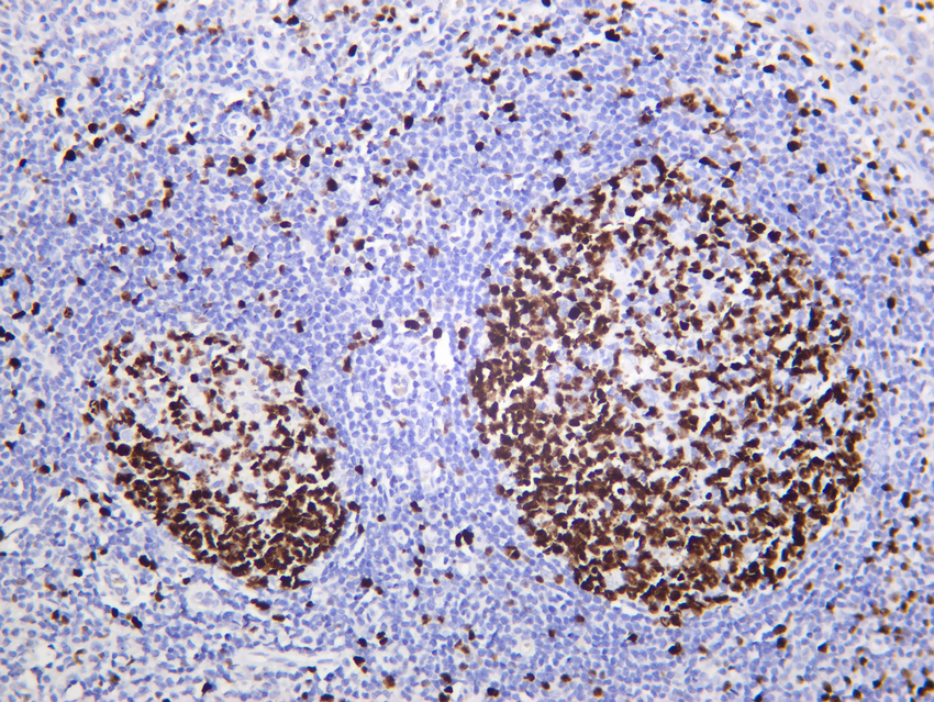
Human tonsil was stained with Anti-Ki67 rabbit antibody.ICC/IF
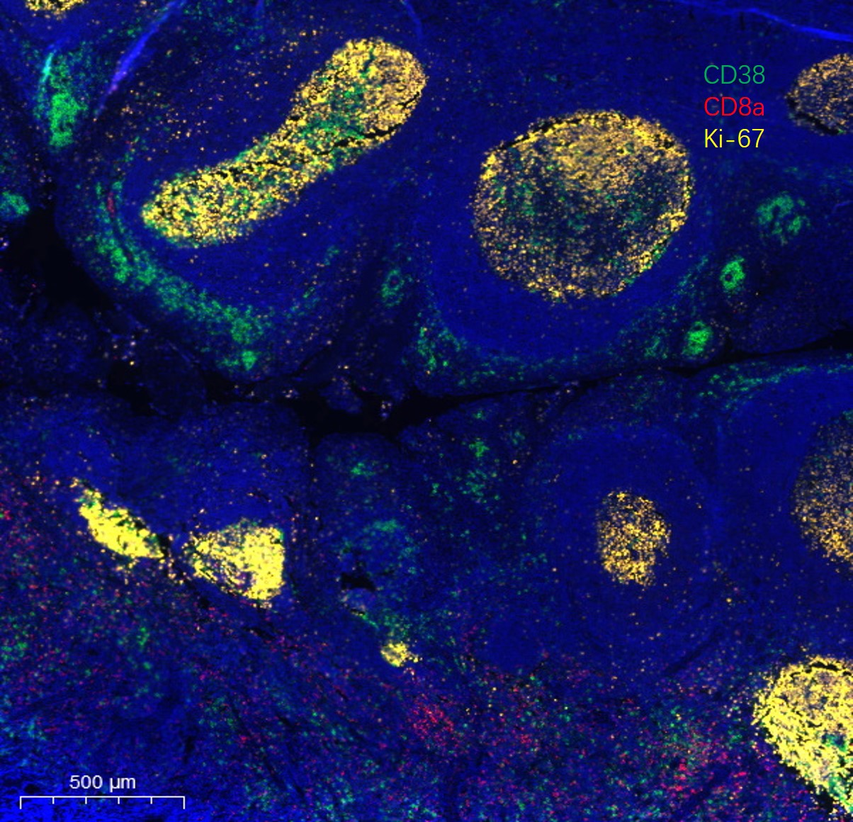
Fluorescence multiplex immunohistochemical analysis of human tonsil tissue (formalin-fixed paraffin-embedded section). The immunostaining was performed by Sextuple-Fluorescence kit (Immunoway).CD8 α rabbit mAbb(RED) and CD38 mouse mAb(GREEN) and Ki67 rabbit mAb(YELLOW) was tested with different TSA Fluorescence regent. Microscopy and pseudocoloring of individual dyes was performed using a Slideviewer Imaging System (Excilone).ICC/IF
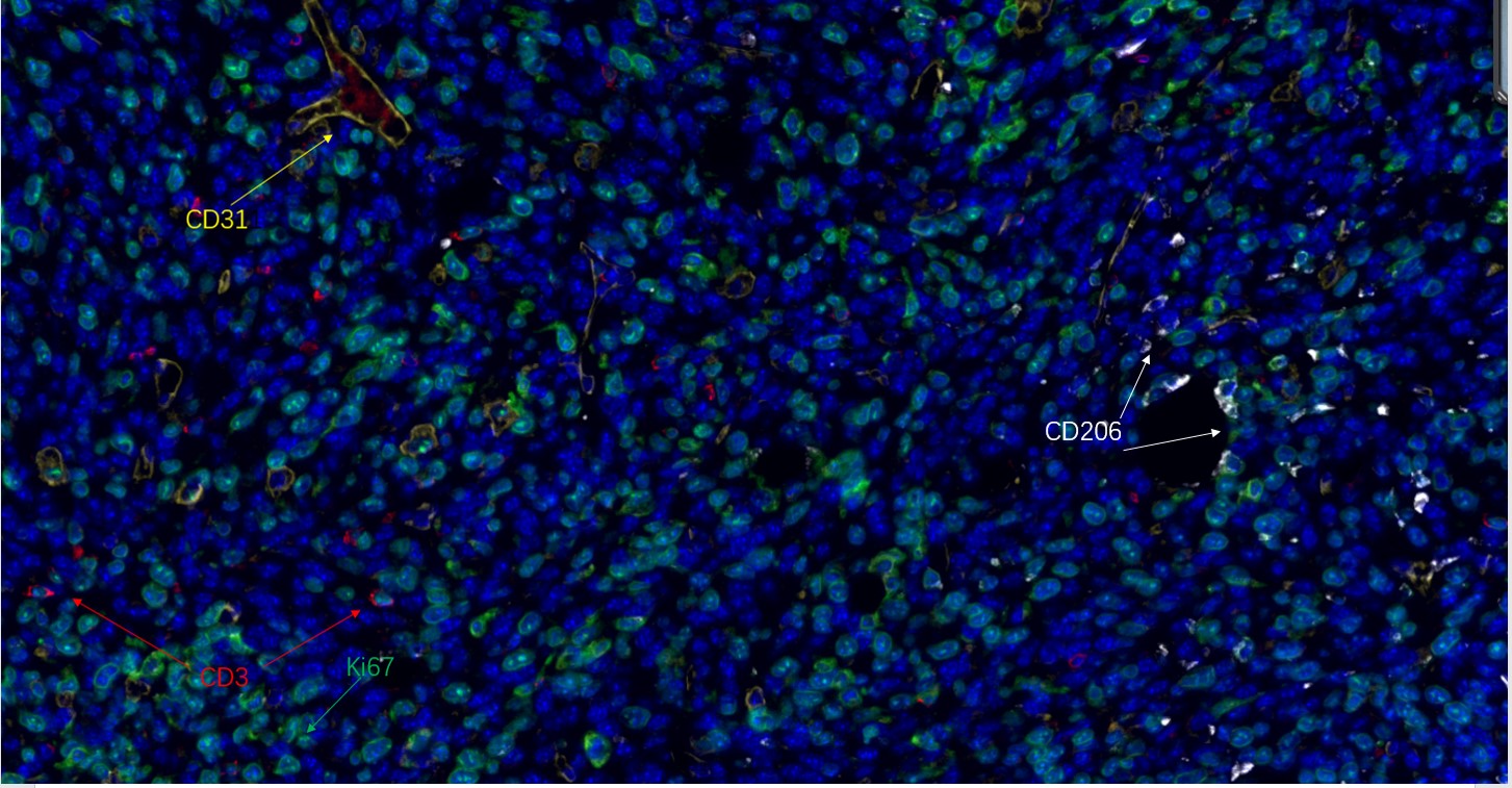
Fluorescence multiplex immunohistochemical analysis of human tonsil tissue (formalin-fixed paraffin-embedded section). The immunostaining was performed by Sextuple-Fluorescence kit (RS0039, Immunoway).CD3 rabbit mAb(RED), Ki67 rabbit mAb(GREEN), CD31 rabbit mAb(YELLOW), CD206 Rabbit mAb(WHITE) was tested with different TSA Fluorescence regent. Microscopy and pseudocoloring of individual dyes was performed using a Slideviewer Imaging System (Excilone).ICC/IF
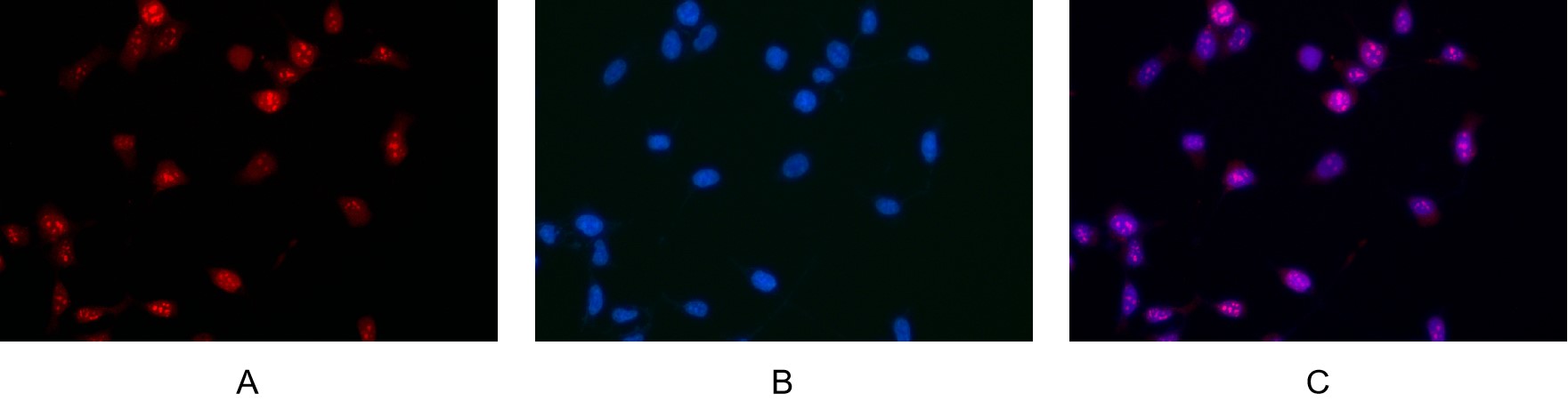
Immunofluorescence analysis of HEK293. Picture A: Ki67 antibody (red). Picture B: DAPI (blue). Picture C: Merge of A+B.| Product Name | Ki-67 Rabbit mAb |
|---|---|
| Antibody Type | Primary Antibodies |
| Clonality | monoclonal |
|---|---|
| Isotype | IgG,K |
| Host Species | Rabbit |
| Tested Applications | ICC/IFIHCWB |
| WB:1:1000-5000 IHC:1:200-1000 ICC/IF:1:200-1:1000 |
|
| Species Reactivity | HumanMouseRat |
| Concentration | 1mg/ml |
| Purification | Protein A |
| Gene Symbol | MKI67 |
|---|---|
| Gene Synonyms | KIA MIB- MIB-1 PPP1R105 |
| Gene Full Name | marker of proliferation Ki-67 |
| Gene Summary | Enables RNA binding activity and molecular condensate scaffold activity. Involved in chromosome segregation and regulation of mitotic nuclear division. Located in chromosome; nucleolus; and nucleoplasm. Is active in condensed chromosome. Implicated in several diseases, including Crohn's disease; colorectal cancer; endocrine gland cancer (multiple); graft-versus-host disease; and human immunodeficiency virus infectious disease. Biomarker of several diseases, including Barrett's esophagus; autoimmune disease of musculoskeletal system (multiple); endocrine gland cancer (multiple); gastrointestinal system cancer (multiple); and lung cancer (multiple). [provided by Alliance of Genome Resources, Jul 2025] |
| Alternative Names | MKI67 Antigen KI-67 |
| Molecular Weight(MW) | 359kDa(Observed MW 359kDa) |
| Cellular Localization | Nucleus |
WB
Various whole cell lysates were separated by 4-8% SDS-PAGE, and the membrane was blotted with anti-Ki67 antibody. The HRP-conjugated Goat anti-Rabbit IgG(H + L) antibody was used to detect the antibody. Lane 1: Hela.
IHC
Rat colon was stained with Anti-Ki67 rabbit antibody.
IHC
Mouse colon was stained with Anti-Ki67 rabbit antibody.
IHC
Human tonsil was stained with Anti-Ki67 rabbit antibody.
ICC/IF
Fluorescence multiplex immunohistochemical analysis of human tonsil tissue (formalin-fixed paraffin-embedded section). The immunostaining was performed by Sextuple-Fluorescence kit (Immunoway).CD8 α rabbit mAbb(RED) and CD38 mouse mAb(GREEN) and Ki67 rabbit mAb(YELLOW) was tested with different TSA Fluorescence regent. Microscopy and pseudocoloring of individual dyes was performed using a Slideviewer Imaging System (Excilone).
ICC/IF
Fluorescence multiplex immunohistochemical analysis of human tonsil tissue (formalin-fixed paraffin-embedded section). The immunostaining was performed by Sextuple-Fluorescence kit (RS0039, Immunoway).CD3 rabbit mAb(RED), Ki67 rabbit mAb(GREEN), CD31 rabbit mAb(YELLOW), CD206 Rabbit mAb(WHITE) was tested with different TSA Fluorescence regent. Microscopy and pseudocoloring of individual dyes was performed using a Slideviewer Imaging System (Excilone).
ICC/IF
Immunofluorescence analysis of HEK293. Picture A: Ki67 antibody (red). Picture B: DAPI (blue). Picture C: Merge of A+B.| Application Notes | WB:1:1000-5000 IHC:1:200-1000 ICC/IF:1:200-1:1000 |
|---|
| Form | Liquid |
|---|---|
| Storage Instructions | -15°C to -25°C/1 year(Do not lower than -25°C) |
| Storage Buffer | PBS, 50% glycerol, 0.05% Proclin 300, 0.05%BSA |


