WB
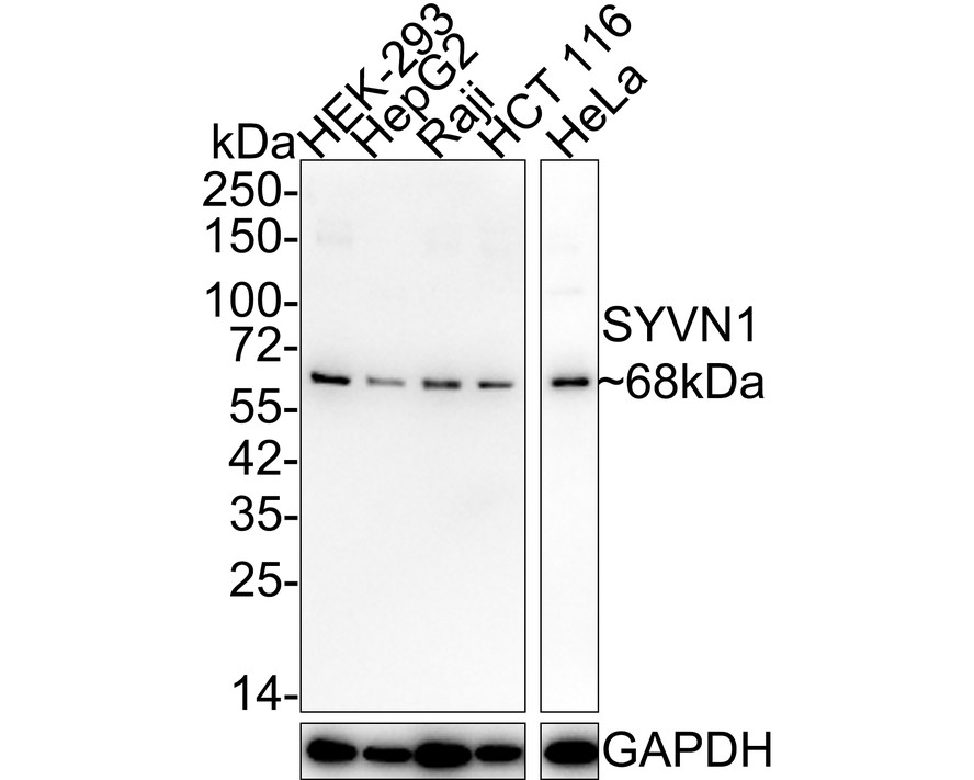
Western blot analysis of SYVN1 on different lysates with Rabbit anti-SYVN1 antibody at 1/2,000 dilution. Lane 1: HEK-293 cell lysate, Lane 2: HepG2 cell lysate, Lane 3: Raji cell lysate, Lane 4: HCT 116 cell lysate, Lane 5: HeLa cell lysate, Lysates/proteins at 20 µg/Lane. Exposure time: 1 minute; 4-20% SDS-PAGE gel. Proteins were transferred to a PVDF membrane and blocked with 5% NFDM/TBST for 1 hour at room temperature. The primary antibody at 1/2,000 dilution was used in 5% NFDM/TBST at 4℃ overnight. Goat Anti-Rabbit IgG - HRP Secondary Antibody at 1/50,000 dilution was used for 1 hour at room temperature.ICC/IF
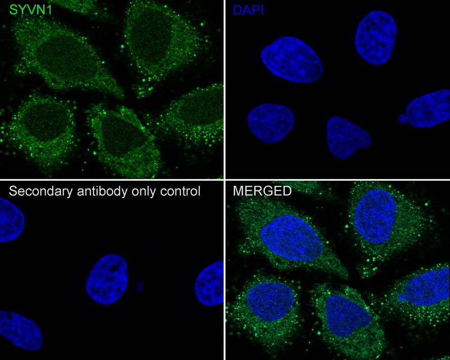
Immunocytochemistry analysis of HeLa cells labeling SYVN1 with Rabbit anti-SYVN1 antibody at 1/100 dilution. Cells were fixed in 4% paraformaldehyde for 20 minutes at room temperature, permeabilized with 0.1% Triton X-100 in PBS for 5 minutes at room temperature, then blocked with 1% BSA in 10% negative goat serum for 1 hour at room temperature. Cells were then incubated with Rabbit anti-SYVN1 antibody at 1/100 dilution in 1% BSA in PBST overnight at 4 ℃. Goat Anti-Rabbit IgG H&L (488) was used as the secondary antibody at 1/1,000 dilution. PBS instead of the primary antibody was used as the secondary antibody only control. Nuclear DNA was labelled in blue with DAPI.FC
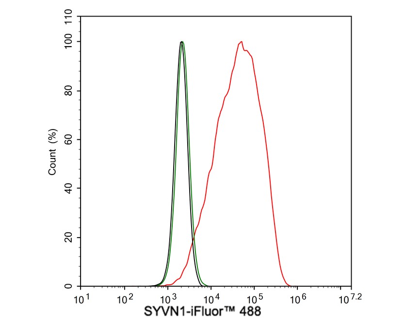
Flow cytometric analysis of HeLa cells labeling SYVN1. Cells were fixed and permeabilized. Then stained with the primary antibody (1μg/mL) (red) compared with Rabbit IgG Isotype Control (green). After incubation of the primary antibody at +4℃ for an hour, the cells were stained with a 488 conjugate-Goat anti-Rabbit IgG Secondary antibody at 1/1,000 dilution for 30 minutes at +4℃. Unlabelled sample was used as a control (cells without incubation with primary antibody; black).| Product Name | SYVN1 Recombinant Rabbit Monoclonal Antibody |
|---|---|
| Antibody Type | Primary Antibodies |
| Immunogen | Synthetic peptide within Human SYVN1 aa 150-250. |
| Clonality | Monoclonal |
|---|---|
| Isotype | IgG |
| Host Species | Rabbit |
| Tested Applications | FCICC/IFWB |
| WB:1:2000 ICC/IF:1:100 FC:1:1000 |
|
| Species Reactivity | Human |
| Concentration | 1mg/ml |
| Purification | Protein A |
| Gene Symbol | SYVN1 |
|---|---|
| Gene Synonyms | DER3 HRD1 |
| Gene Full Name | synoviolin 1 |
| Gene Summary | This gene encodes a protein involved in endoplasmic reticulum (ER)-associated degradation. The encoded protein removes unfolded proteins, accumulated during ER stress, by retrograde transport to the cytosol from the ER. This protein also uses the ubiquitin-proteasome system for additional degradation of unfolded proteins. Sequence analysis identified two transcript variants that encode different isoforms. [provided by RefSeq, May 2011] |
| Alternative Names | 1200010C09Rik antibody DER3 antibody E3 ubiquitin-protein ligase synoviolin antibody HMG coA reductase degradation 1 homolog antibody HRD1 antibody KIAA1810 antibody MGC40372 antibody OTTHUMP00000230429 antibody OTTHUMP00000230430 antibody OTTHUMP00000230431 antibody 1200010C09Rik antibody DER3 antibody E3 ubiquitin-protein ligase synoviolin antibody HMG coA reductase degradation 1 homolog antibody HRD1 antibody KIAA1810 antibody MGC40372 antibody OTTHUMP00000230429 antibody OTTHUMP00000230430 antibody OTTHUMP00000230431 antibody OTTHUMP00000230432 antibody Synovial apoptosis inhibitor 1 antibody Synovial apoptosis inhibitor 1 synoviolin antibody Synoviolin 1 antibody Synoviolin 1 isoform b antibody SYNOVIOLIN antibody SYVN1 antibody SYVN1_HUMAN antibody |
| Molecular Weight(MW) | 68 kDa |
| Cellular Localization | Endoplasmic reticulum membrane. |
WB
Western blot analysis of SYVN1 on different lysates with Rabbit anti-SYVN1 antibody at 1/2,000 dilution. Lane 1: HEK-293 cell lysate, Lane 2: HepG2 cell lysate, Lane 3: Raji cell lysate, Lane 4: HCT 116 cell lysate, Lane 5: HeLa cell lysate, Lysates/proteins at 20 µg/Lane. Exposure time: 1 minute; 4-20% SDS-PAGE gel. Proteins were transferred to a PVDF membrane and blocked with 5% NFDM/TBST for 1 hour at room temperature. The primary antibody at 1/2,000 dilution was used in 5% NFDM/TBST at 4℃ overnight. Goat Anti-Rabbit IgG - HRP Secondary Antibody at 1/50,000 dilution was used for 1 hour at room temperature.
ICC/IF
Immunocytochemistry analysis of HeLa cells labeling SYVN1 with Rabbit anti-SYVN1 antibody at 1/100 dilution. Cells were fixed in 4% paraformaldehyde for 20 minutes at room temperature, permeabilized with 0.1% Triton X-100 in PBS for 5 minutes at room temperature, then blocked with 1% BSA in 10% negative goat serum for 1 hour at room temperature. Cells were then incubated with Rabbit anti-SYVN1 antibody at 1/100 dilution in 1% BSA in PBST overnight at 4 ℃. Goat Anti-Rabbit IgG H&L (488) was used as the secondary antibody at 1/1,000 dilution. PBS instead of the primary antibody was used as the secondary antibody only control. Nuclear DNA was labelled in blue with DAPI.
FC
Flow cytometric analysis of HeLa cells labeling SYVN1. Cells were fixed and permeabilized. Then stained with the primary antibody (1μg/mL) (red) compared with Rabbit IgG Isotype Control (green). After incubation of the primary antibody at +4℃ for an hour, the cells were stained with a 488 conjugate-Goat anti-Rabbit IgG Secondary antibody at 1/1,000 dilution for 30 minutes at +4℃. Unlabelled sample was used as a control (cells without incubation with primary antibody; black).| Application Notes | WB:1:2000 ICC/IF:1:100 FC:1:1000 |
|---|
| Form | Liquid |
|---|---|
| Storage Instructions | Store at +4℃ after thawing. Aliquot store at -20℃. Avoid repeated freeze / thaw cycles. |
| Storage Buffer | 1*TBS (pH7.4), 0.05% BSA, 40% Glycerol. Preservative: 0.05% Sodium Azide. |


