WB
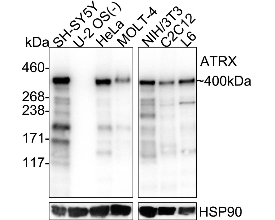
Western blot analysis of ATRX on different lysates with Rabbit anti-ATRX antibody at 1/5,000 dilution. Lane 1: SH-SY5Y cell lysate Lane 2: U-2 OS cell lysate (negative) Lane 3: HeLa cell lysate Lane 4: MOLT-4 cell lysate Lane 5: NIH/3T3 cell lysate Lane 6: C2C12 cell lysate Lane 7: L6 cell lysate Lysates/proteins at 20 µg/Lane. Exposure time: 10 seconds; 3-8% SDS-PAGE gel. Proteins were transferred to a PVDF membrane and blocked with 5% NFDM/TBST for 1 hour at room temperature. The primary antibody at 1/5,000 dilution was used in primary antibody dilution at 4℃ overnight. Goat Anti-Rabbit IgG - HRP Secondary Antibody at 1/50,000 dilution was used for 1 hour at room temperature.IHC
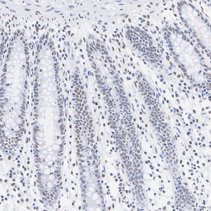
Immunohistochemical analysis of paraffin-embedded human colon tissue with Rabbit anti-ATRX antibody at 1/200 dilution. The section was pre-treated using heat mediated antigen retrieval with Tris-EDTA buffer (pH 9.0) for 20 minutes. The tissues were blocked in 1% BSA for 20 minutes at room temperature, washed with ddH2O and PBS, and then probed with the primary antibody at 1/200 dilution for 1 hour at room temperature. The detection was performed using an HRP conjugated compact polymer system. DAB was used as the chromogen. Tissues were counterstained with hematoxylin and mounted with DPX.IHC
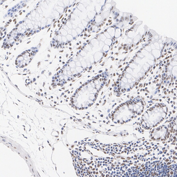
Immunohistochemical analysis of paraffin-embedded mouse colon tissue with Rabbit anti-ATRX antibody at 1/200 dilution. The section was pre-treated using heat mediated antigen retrieval with Tris-EDTA buffer (pH 9.0) for 20 minutes. The tissues were blocked in 1% BSA for 20 minutes at room temperature, washed with ddH2O and PBS, and then probed with the primary antibody at 1/200 dilution for 1 hour at room temperature. The detection was performed using an HRP conjugated compact polymer system. DAB was used as the chromogen. Tissues were counterstained with hematoxylin and mounted with DPX.IHC
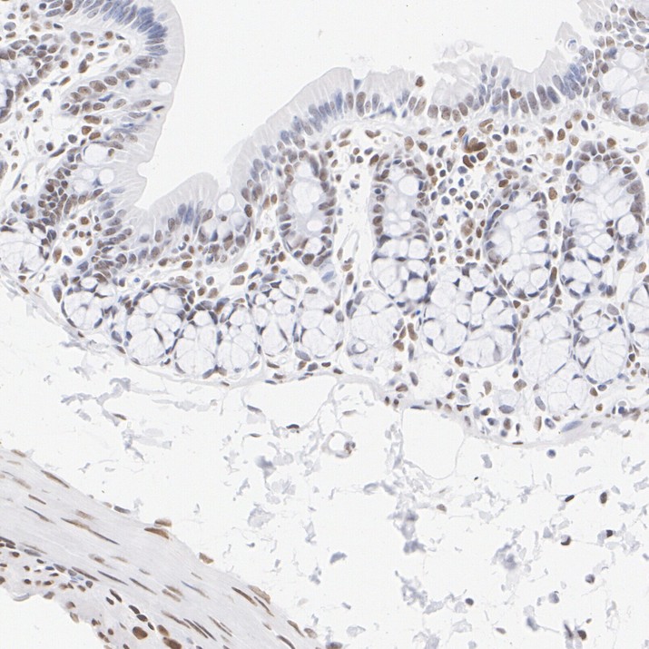
Immunohistochemical analysis of paraffin-embedded rat colon tissue with Rabbit anti-ATRX antibody at 1/200 dilution. The section was pre-treated using heat mediated antigen retrieval with Tris-EDTA buffer (pH 9.0) for 20 minutes. The tissues were blocked in 1% BSA for 20 minutes at room temperature, washed with ddH2O and PBS, and then probed with the primary antibody at 1/200 dilution for 1 hour at room temperature. The detection was performed using an HRP conjugated compact polymer system. DAB was used as the chromogen. Tissues were counterstained with hematoxylin and mounted with DPX.ICC/IF
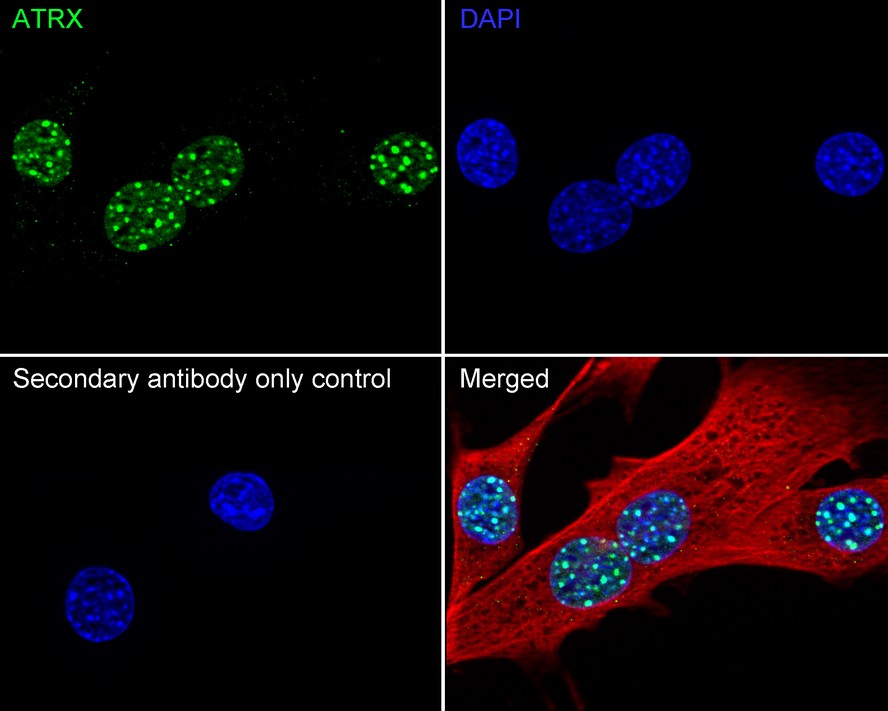
Immunocytochemistry analysis of NIH/3T3 cells labeling ATRX with Rabbit anti-ATRX antibody at 1/1,000 dilution. Cells were fixed in 4% paraformaldehyde for 15 minutes at room temperature, permeabilized with 0.1% Triton X-100 in PBS for 15 minutes at room temperature, then blocked with 1% BSA in 10% negative goat serum for 1 hour at room temperature. Cells were then incubated with Rabbit anti-ATRX antibody at 1/1,000 dilution in 1% BSA in PBST overnight at 4 ℃. Goat Anti-Rabbit IgG H&L (488) was used as the secondary antibody at 1/1,000 dilution. PBS instead of the primary antibody was used as the secondary antibody only control. Nuclear DNA was labelled in blue with DAPI. Beta tubulin (red) was stained at 1/100 dilution overnight at +4℃. Goat Anti-Mouse IgG H&L (594) was used as the secondary antibody at 1/1,000 dilution.IP
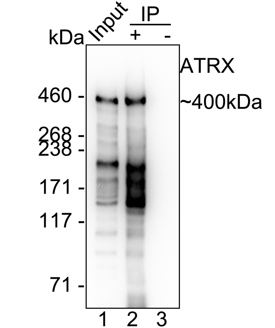
ATRX was immunoprecipitated from 0.2 mg SH-SY5Y cell lysate with Rabbit anti-ATRX antibody at 2 µg/10 µl beads. Western blot was performed from the immunoprecipitate using Rabbit anti-ATRX antibody at 1/5,000 dilution. HRP Conjugated Anti-Rabbit IgG for IP Nano-secondary antibody at 1/5,000 dilution was used for 1 hour at room temperature. Lane 1: SH-SY5Y cell lysate (input), Lane 2: Rabbit anti-ATRX antibody IP in SH-SY5Y cell lysate, Lane 3: Rabbit IgG instead of Rabbit anti-ATRX antibody in SH-SY5Y cell lysate, Blocking/Dilution buffer: primary antibody dilution, Exposure time: 20 seconds.| Product Name | ATRX Recombinant Rabbit Monoclonal Antibody |
|---|---|
| Antibody Type | Primary Antibodies |
| Immunogen | Recombinant protein within human ATRX aa 2,193-2,492. |
| Clonality | Monoclonal |
|---|---|
| Isotype | IgG |
| Host Species | Rabbit |
| Tested Applications | ICC/IFIHCIPWB |
| WB:1:5000-1:10000 IHC:1:200-1:1000 ICC/IF:1:1000 IP:1-2μg/sample |
|
| Species Reactivity | HumanMouseRat |
| Concentration | 1mg/ml |
| Purification | Protein A |
| Gene Symbol | ATRX |
|---|---|
| Gene Synonyms | JMS XH2 XNP MRX52 RAD54 RAD54L ZNF-HX |
| Gene Full Name | ATRX chromatin remodeler |
| Gene Summary | The protein encoded by this gene contains an ATPase/helicase domain, and thus it belongs to the SWI/SNF family of chromatin remodeling proteins. This protein is found to undergo cell cycle-dependent phosphorylation, which regulates its nuclear matrix and chromatin association, and suggests its involvement in the gene regulation at interphase and chromosomal segregation in mitosis. Mutations in this gene are associated with X-linked syndromes exhibiting cognitive disabilities as well as alpha-thalassemia (ATRX) syndrome. These mutations have been shown to cause diverse changes in the pattern of DNA methylation, which may provide a link between chromatin remodeling, DNA methylation, and gene expression in developmental processes. Multiple alternatively spliced transcript variants encoding distinct isoforms have been reported. [provided by RefSeq, Jul 2017] |
| Alternative Names | Alpha thalassemia/mental retardation syndrome X linked homolog antibody ATP dependent helicase ATRX antibody ATP-dependent helicase ATRX antibody ATR2 antibody Atrx antibody ATRX_HUMAN antibody DNA dependent ATPase and helicase antibody Helicase 2 X linked antibody MGC2094 antibody MRXHF1 antibody Alpha thalassemia/mental retardation syndrome X linked homolog antibody ATP dependent helicase ATRX antibody ATP-dependent helicase ATRX antibody ATR2 antibody Atrx antibody ATRX_HUMAN antibody DNA dependent ATPase and helicase antibody Helicase 2 X linked antibody MGC2094 antibody MRXHF1 antibody RAD54 antibody RAD54L antibody SFM1 antibody SHS antibody Transcriptional regulator ATRX antibody X linked helicase II antibody X linked nuclear protein antibody X-linked helicase II antibody X-linked nuclear protein antibody XH2 antibody XNP antibody Znf HX antibody Znf-HX antibody |
| Molecular Weight(MW) | 283 kDa(Observed band size: 400 kDa) |
| Cellular Localization | Nucleus, Chromosome, telomere, PML body. |
WB
Western blot analysis of ATRX on different lysates with Rabbit anti-ATRX antibody at 1/5,000 dilution. Lane 1: SH-SY5Y cell lysate Lane 2: U-2 OS cell lysate (negative) Lane 3: HeLa cell lysate Lane 4: MOLT-4 cell lysate Lane 5: NIH/3T3 cell lysate Lane 6: C2C12 cell lysate Lane 7: L6 cell lysate Lysates/proteins at 20 µg/Lane. Exposure time: 10 seconds; 3-8% SDS-PAGE gel. Proteins were transferred to a PVDF membrane and blocked with 5% NFDM/TBST for 1 hour at room temperature. The primary antibody at 1/5,000 dilution was used in primary antibody dilution at 4℃ overnight. Goat Anti-Rabbit IgG - HRP Secondary Antibody at 1/50,000 dilution was used for 1 hour at room temperature.
IHC
Immunohistochemical analysis of paraffin-embedded human colon tissue with Rabbit anti-ATRX antibody at 1/200 dilution. The section was pre-treated using heat mediated antigen retrieval with Tris-EDTA buffer (pH 9.0) for 20 minutes. The tissues were blocked in 1% BSA for 20 minutes at room temperature, washed with ddH2O and PBS, and then probed with the primary antibody at 1/200 dilution for 1 hour at room temperature. The detection was performed using an HRP conjugated compact polymer system. DAB was used as the chromogen. Tissues were counterstained with hematoxylin and mounted with DPX.
IHC
Immunohistochemical analysis of paraffin-embedded mouse colon tissue with Rabbit anti-ATRX antibody at 1/200 dilution. The section was pre-treated using heat mediated antigen retrieval with Tris-EDTA buffer (pH 9.0) for 20 minutes. The tissues were blocked in 1% BSA for 20 minutes at room temperature, washed with ddH2O and PBS, and then probed with the primary antibody at 1/200 dilution for 1 hour at room temperature. The detection was performed using an HRP conjugated compact polymer system. DAB was used as the chromogen. Tissues were counterstained with hematoxylin and mounted with DPX.
IHC
Immunohistochemical analysis of paraffin-embedded rat colon tissue with Rabbit anti-ATRX antibody at 1/200 dilution. The section was pre-treated using heat mediated antigen retrieval with Tris-EDTA buffer (pH 9.0) for 20 minutes. The tissues were blocked in 1% BSA for 20 minutes at room temperature, washed with ddH2O and PBS, and then probed with the primary antibody at 1/200 dilution for 1 hour at room temperature. The detection was performed using an HRP conjugated compact polymer system. DAB was used as the chromogen. Tissues were counterstained with hematoxylin and mounted with DPX.
ICC/IF
Immunocytochemistry analysis of NIH/3T3 cells labeling ATRX with Rabbit anti-ATRX antibody at 1/1,000 dilution. Cells were fixed in 4% paraformaldehyde for 15 minutes at room temperature, permeabilized with 0.1% Triton X-100 in PBS for 15 minutes at room temperature, then blocked with 1% BSA in 10% negative goat serum for 1 hour at room temperature. Cells were then incubated with Rabbit anti-ATRX antibody at 1/1,000 dilution in 1% BSA in PBST overnight at 4 ℃. Goat Anti-Rabbit IgG H&L (488) was used as the secondary antibody at 1/1,000 dilution. PBS instead of the primary antibody was used as the secondary antibody only control. Nuclear DNA was labelled in blue with DAPI. Beta tubulin (red) was stained at 1/100 dilution overnight at +4℃. Goat Anti-Mouse IgG H&L (594) was used as the secondary antibody at 1/1,000 dilution.
IP
ATRX was immunoprecipitated from 0.2 mg SH-SY5Y cell lysate with Rabbit anti-ATRX antibody at 2 µg/10 µl beads. Western blot was performed from the immunoprecipitate using Rabbit anti-ATRX antibody at 1/5,000 dilution. HRP Conjugated Anti-Rabbit IgG for IP Nano-secondary antibody at 1/5,000 dilution was used for 1 hour at room temperature. Lane 1: SH-SY5Y cell lysate (input), Lane 2: Rabbit anti-ATRX antibody IP in SH-SY5Y cell lysate, Lane 3: Rabbit IgG instead of Rabbit anti-ATRX antibody in SH-SY5Y cell lysate, Blocking/Dilution buffer: primary antibody dilution, Exposure time: 20 seconds.| Application Notes | WB:1:5000-1:10000 IHC:1:200-1:1000 ICC/IF:1:1000 IP:1-2μg/sample |
|---|
| Form | Liquid |
|---|---|
| Storage Instructions | Store at +4℃ after thawing. Aliquot store at -20℃. Avoid repeated freeze / thaw cycles. |
| Storage Buffer | 1*TBS (pH7.4), 0.05% BSA, 40% Glycerol. Preservative: 0.05% Sodium Azide. |


