WB
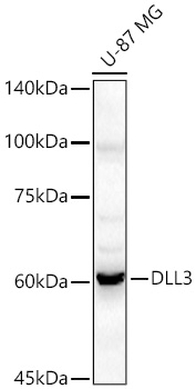
Western blot analysis of various lysates using DLL3 Rabbit pAb at 1:500 dilution. Secondary antibody: HRP-conjugated Goat anti-Rabbit IgG (H+L) at 1:10000 dilution. Lysates/proteins: 25μg per lane. Blocking buffer: 3% nonfat dry milk in TBST. Detection: ECL Basic Kit. Exposure time: 90s.IHC
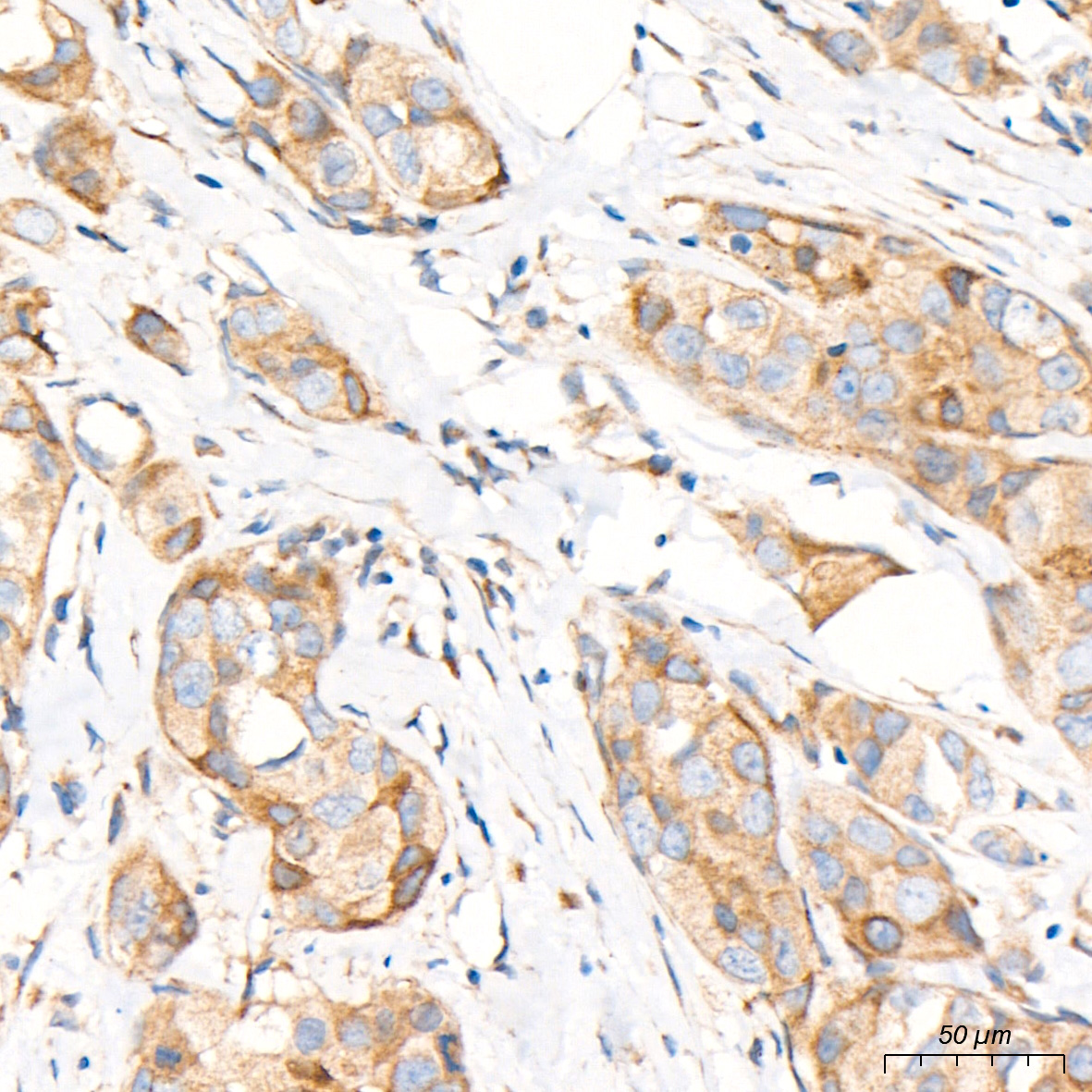
Immunohistochemistry analysis of paraffin embedded Human breast cancer tissue using DLL3 Rabbit pAb at a dilution of 1:200 (40x lens). High pressure antigen retrieval performed with 0.01M Tris-EDTA Buffer (pH 9.0) prior to IHC staining.IHC
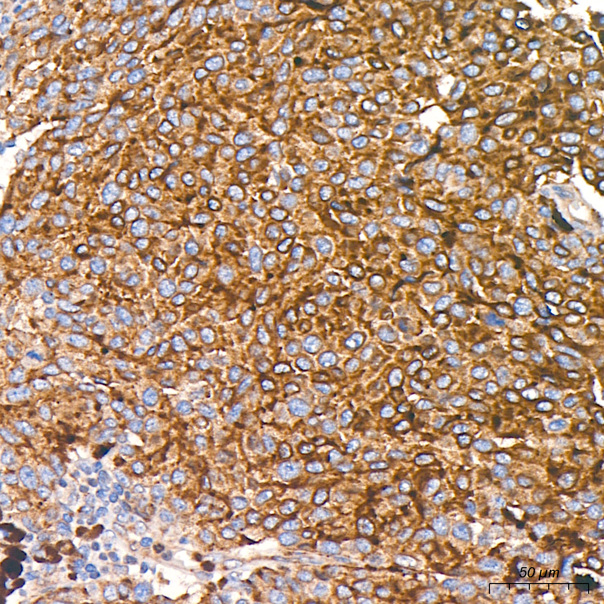
Immunohistochemistry analysis of paraffin embedded Human malignant melanoma tissue using DLL3 Rabbit pAb at a dilution of 1:200 (40x lens). High pressure antigen retrieval performed with 0.01M Tris EDTA Buffer (pH 9.0) prior to IHC staining.IHC
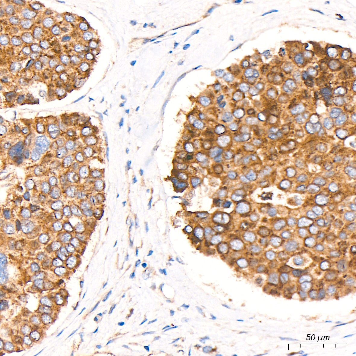
Immunohistochemistry analysis of paraffin embedded Human pancreatic cancer tissue using DLL3 Rabbit pAb at a dilution of 1:200 (40x lens). High pressure antigen retrieval performed with 0.01M Tris-EDTA Buffer (pH 9.0) prior to IHC staining.| Product Name | DLL3 Rabbit pAb |
|---|---|
| Antibody Type | Primary Antibodies |
| Immunogen | Recombinant protein. |
| Clonality | Polyclonal |
|---|---|
| Isotype | IgG |
| Host Species | Rabbit |
| Tested Applications | IHCWB |
| WB:1:100-1:500 IHC:1:100-1:400 |
|
| Species Reactivity | HumanMouseRat |
| Concentration | 1mg/ml |
| Purification | Affinity purified |
| Gene Symbol | DLL3 |
|---|---|
| Gene Synonyms | SCDO1 |
| Gene Full Name | delta like canonical Notch ligand 3 |
| Gene Summary | This gene encodes a member of the delta protein ligand family. This family functions as Notch ligands that are characterized by a DSL domain, EGF repeats, and a transmembrane domain. Mutations in this gene cause autosomal recessive spondylocostal dysostosis 1. Two transcript variants encoding distinct isoforms have been identified for this gene. [provided by RefSeq, Jul 2008] |
| Alternative Names | SCDO1 DLL3 |
| Molecular Weight(MW) | 65 kDa |
| Cellular Localization | Membrane. |
WB
Western blot analysis of various lysates using DLL3 Rabbit pAb at 1:500 dilution. Secondary antibody: HRP-conjugated Goat anti-Rabbit IgG (H+L) at 1:10000 dilution. Lysates/proteins: 25μg per lane. Blocking buffer: 3% nonfat dry milk in TBST. Detection: ECL Basic Kit. Exposure time: 90s.
IHC
Immunohistochemistry analysis of paraffin embedded Human breast cancer tissue using DLL3 Rabbit pAb at a dilution of 1:200 (40x lens). High pressure antigen retrieval performed with 0.01M Tris-EDTA Buffer (pH 9.0) prior to IHC staining.
IHC
Immunohistochemistry analysis of paraffin embedded Human malignant melanoma tissue using DLL3 Rabbit pAb at a dilution of 1:200 (40x lens). High pressure antigen retrieval performed with 0.01M Tris EDTA Buffer (pH 9.0) prior to IHC staining.
IHC
Immunohistochemistry analysis of paraffin embedded Human pancreatic cancer tissue using DLL3 Rabbit pAb at a dilution of 1:200 (40x lens). High pressure antigen retrieval performed with 0.01M Tris-EDTA Buffer (pH 9.0) prior to IHC staining.| Application Notes | WB:1:100-1:500 IHC:1:100-1:400 |
|---|
| Form | Liquid |
|---|---|
| Storage Instructions | Store at -20℃. Avoid freeze / thaw cycles. |
| Storage Buffer | Buffer: PBS with 0.05% proclin300, 0.05% BSA, 50% glycerol, pH7.3. |


