WB
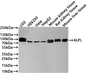
Positive WB detected in: LO2 whole cell lysate(30µg), HEK293 whole cell lysate(30µg), Hela whole cell lysate(30µg), HepG2 whole cell lysate(30µg), Mouse kidney tissue lysate(20µg), Rat kidney tissue lysate(20µg), Mouse liver tissue lysate(20µg). All lanes: ALPL antibody at 1:1000. Secondary Goat polyclonal to rabbit IgG at 1/40000 dilution.Exposure time:2min.IHC
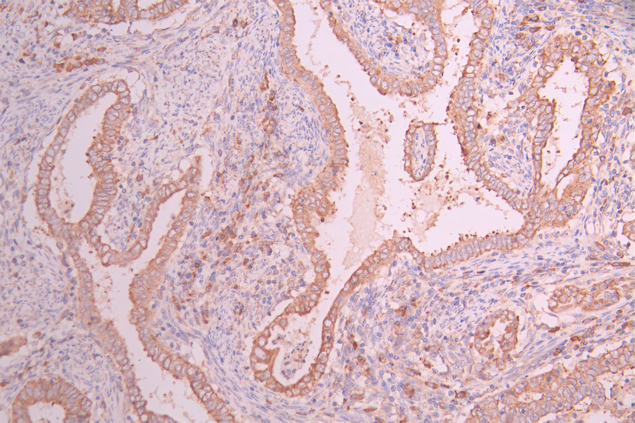
IHC image of ALPL Recombinant Monoclonal Antibody diluted at 1:100 and staining in paraffin-embedded human endometrial cancer/endometrial carcinoma performed on a Leica BondTM system. After dewaxing and hydration, antigen retrieval was mediated by high pressure in a citrate buffer (pH 6.0). Section was blocked with 10% normal goat serum 30min at RT. Then primary antibody (1% BSA) was incubated at 4°C overnight. The primary is detected by a Goat anti-rabbit polymer IgG labeled by HRP and visualized using 0.05% DAB.IHC
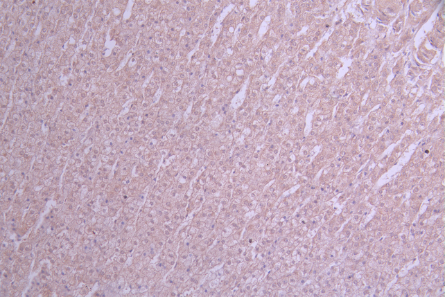
IHC image of ALPL Recombinant Monoclonal Antibody diluted at 1:100 and staining in paraffin-embedded human adrenal gland tissue performed on a Leica BondTM system. After dewaxing and hydration, antigen retrieval was mediated by high pressure in a citrate buffer (pH 6.0). Section was blocked with 10% normal goat serum 30min at RT. Then primary antibody (1% BSA) was incubated at 4°C overnight. The primary is detected by a Goat anti-rabbit polymer IgG labeled by HRP and visualized using 0.05% DAB.ICC/IF
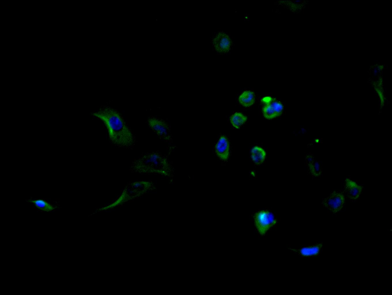
Immunofluorescence staining of HepG2 cell with ALPL Recombinant Monoclonal Antibody at 1:50, counter-stained with DAPI. The cells were fixed in 4% formaldehyde and blocked in 10% normal Goat Serum. The cells were then incubated with the antibody overnight at 4°C. The secondary antibody was Alexa Fluor 488-congugated AffiniPure Goat Anti-Rabbit IgG(H+L).FC
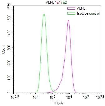
Overlay Peak curve showing Hela cells stained with ALPL Recombinant Monoclonal Antibody (red line) at 1:100. The cells were fixed in 4% formaldehyde and permeated by 0.2% TritonX-100 for 10min. Then 10% normal goat serum to block non-specific protein-protein interactions followed by the antibody (1ug/1*106cells) for 45min at 4℃. The secondary antibody used was FITC-conjugated goat anti-rabbit IgG (H+L) at 1/200 dilution for 35min at 4℃.Control antibody (green line) was Rabbit IgG (1ug/1*106cells) used under the same conditions. Acquisition of >10,000 events was performed.| Product Name | ALPL Recombinant Monoclonal Antibody |
|---|---|
| Antibody Type | Primary Antibodies |
| Immunogen | A synthesized peptide derived from human ALPL |
| Clonality | Monoclonal |
|---|---|
| Isotype | IgG |
| Host Species | Rabbit |
| Tested Applications | FCICC/IFIHCWB |
| WB:1:500-1:2000 IHC:1:50-1:200 ICC/IF:1:50-1:200 FC:1:50-1:200 |
|
| Species Reactivity | HumanMouseRat |
| Concentration | 1mg/ml |
| Purification | Affinity purified |
| Gene Symbol | ALPL |
|---|---|
| Gene Synonyms | HOPS HPPA HPPC HPPI HPPO TNAP TNALP APTNAP TNSALP AP-TNAP TNS-ALP |
| Gene Full Name | alkaline phosphatase, biomineralization associated |
| Gene Summary | This gene encodes a member of the alkaline phosphatase family of proteins. There are at least four distinct but related alkaline phosphatases: intestinal, placental, placental-like, and liver/bone/kidney (tissue non-specific). The first three are located together on chromosome 2, while the tissue non-specific form is located on chromosome 1. The product of this gene is a membrane bound glycosylated enzyme that is not expressed in any particular tissue and is, therefore, referred to as the tissue-nonspecific form of the enzyme. Alternative splicing results in multiple transcript variants, at least one of which encodes a preproprotein that is proteolytically processed to generate the mature enzyme. This enzyme may play a role in bone mineralization. Mutations in this gene have been linked to hypophosphatasia, a disorder that is characterized by hypercalcemia and skeletal defects. [provided by RefSeq, Oct 2015] |
| Alternative Names | AKP2 antibody Alkaline phosphatase liver/bone/kidney antibody Alkaline phosphatase liver/bone/kidney isozyme antibody Alkaline phosphatase tissue nonspecific isozyme antibody Alkaline phosphatase tissue-nonspecific isozyme antibody Alkaline phosphomonoesterase antibody Alpl antibody AP TNAP antibody AP-TNAP antibody APTNAP antibody BAP antibody FLJ40094 antibody FLJ93059 antibody Glycerophosphatase antibody HOPS antibody Liver/bone/kidney type alkaline phosphatase antibody MGC161443 antibody MGC167935 antibody PHOA antibody PPBT_HUMAN antibody Tissue non specific alkaline phosphatase antibody Tissue nonspecific ALP antibody TNAP antibody TNSALP antibody |
| Molecular Weight(MW) | 57 kDa(Observed band size: 75 kDa) |
| Cellular Localization | Cell membrane; Lipid-anchor, GPI-anchor. Extracellular vesicle membrane; Lipid-anchor, GPI-anchor. Mitochondrion membrane; Lipid-anchor, GPI-anchor. Mitochondrion intermembrane space. |
WB
Positive WB detected in: LO2 whole cell lysate(30µg), HEK293 whole cell lysate(30µg), Hela whole cell lysate(30µg), HepG2 whole cell lysate(30µg), Mouse kidney tissue lysate(20µg), Rat kidney tissue lysate(20µg), Mouse liver tissue lysate(20µg). All lanes: ALPL antibody at 1:1000. Secondary Goat polyclonal to rabbit IgG at 1/40000 dilution.Exposure time:2min.
IHC
IHC image of ALPL Recombinant Monoclonal Antibody diluted at 1:100 and staining in paraffin-embedded human endometrial cancer/endometrial carcinoma performed on a Leica BondTM system. After dewaxing and hydration, antigen retrieval was mediated by high pressure in a citrate buffer (pH 6.0). Section was blocked with 10% normal goat serum 30min at RT. Then primary antibody (1% BSA) was incubated at 4°C overnight. The primary is detected by a Goat anti-rabbit polymer IgG labeled by HRP and visualized using 0.05% DAB.
IHC
IHC image of ALPL Recombinant Monoclonal Antibody diluted at 1:100 and staining in paraffin-embedded human adrenal gland tissue performed on a Leica BondTM system. After dewaxing and hydration, antigen retrieval was mediated by high pressure in a citrate buffer (pH 6.0). Section was blocked with 10% normal goat serum 30min at RT. Then primary antibody (1% BSA) was incubated at 4°C overnight. The primary is detected by a Goat anti-rabbit polymer IgG labeled by HRP and visualized using 0.05% DAB.
ICC/IF
Immunofluorescence staining of HepG2 cell with ALPL Recombinant Monoclonal Antibody at 1:50, counter-stained with DAPI. The cells were fixed in 4% formaldehyde and blocked in 10% normal Goat Serum. The cells were then incubated with the antibody overnight at 4°C. The secondary antibody was Alexa Fluor 488-congugated AffiniPure Goat Anti-Rabbit IgG(H+L).
FC
Overlay Peak curve showing Hela cells stained with ALPL Recombinant Monoclonal Antibody (red line) at 1:100. The cells were fixed in 4% formaldehyde and permeated by 0.2% TritonX-100 for 10min. Then 10% normal goat serum to block non-specific protein-protein interactions followed by the antibody (1ug/1*106cells) for 45min at 4℃. The secondary antibody used was FITC-conjugated goat anti-rabbit IgG (H+L) at 1/200 dilution for 35min at 4℃.Control antibody (green line) was Rabbit IgG (1ug/1*106cells) used under the same conditions. Acquisition of >10,000 events was performed.| Application Notes | WB:1:500-1:2000 IHC:1:50-1:200 ICC/IF:1:50-1:200 FC:1:50-1:200 |
|---|
| Form | Liquid |
|---|---|
| Storage Instructions | Upon receipt, store at -20°C or -80°C. Avoid repeated freeze. |
| Storage Buffer | 0.03% Proclin 300 Constituents: 50% Glycerol, 0.01M PBS, PH 7.4 |


